By Marcello Cherchi, MD PhD
For patients
Your doctor may check your balance with a modified version of something called the Romberg test. In this test you will stand with one foot in front of the other, touching heel to toes, with your eyes closed. Most individuals can maintain this stance for some seconds before falling (or breaking out of the stance to prevent a fall). The shorter the time that you can maintain this position, the more likely it is that you have a balance problem, though this does not by itself tell your doctor what the underlying cause of the imbalance is. Depending on this result, your doctor may consider checking other tests of balance function. The Romberg test is not uncomfortable. It takes about 20 – 30 seconds to perform. You do not need any special preparation for this test. You do not need to do anything specific after the test.
For clinicians
Overview
In its original formulation, the Romberg test was used as a test for lower extremity somatosensory deficits from tabes dorsalis (spinal cord posterior column dysfunction secondary to neurosyphilis). It is sometimes used in a modified form in otoneurology by having a patient close their eyes while in unsighted tandem Romberg stance. The less time that passes before the patient falls (or breaks out of the stance in order to prevent a fall), the more likely it is that there is a deficit in proprioception and/or vestibular function. The time to fall also becomes shorter with normal aging due to presbyvestibulopathy. The test is brief and easy to perform, and it is sensitive but not specific.
Introduction
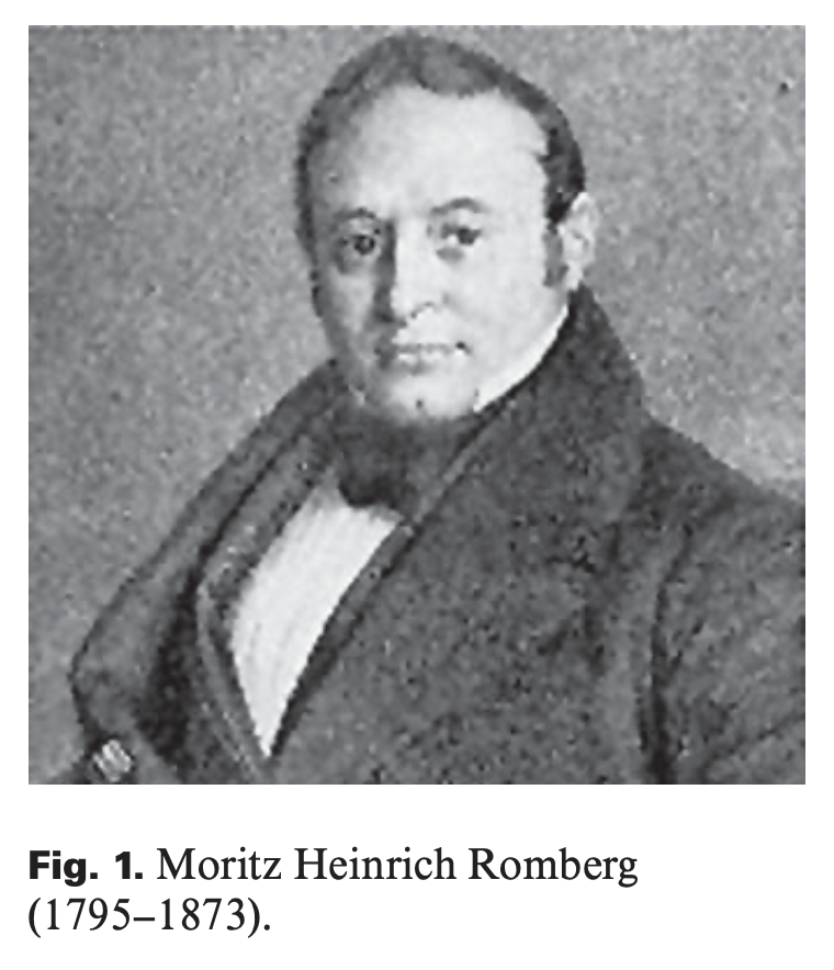
Moritz Heinrich Romberg (1795 – 1873) studied medicine in Berlin and Vienna, and then returned to Berlin where he remained for the rest of his career (Pearce 2005).
Among his many neurological interests, Romberg is remembered primarily for his study of patients with tabes dorsalis (degeneration of the posterior columns of the spinal cord leading to reduced pallesthesia and proprioception in the lower extremities, causing progressive sensory “locomotor ataxia”), which was later recognized to be a manifestation of neurosyphilis (Housman et al. 2014). In the first edition of Romberg’s 3-volume manual of neurology (Romberg 1846) he stated (as translated by Edward Sieveking):
“If the patient [with tabes dorsalis] is told to shut his eyes while in the erect posture, he immediately begins to move from side to side, and the oscillations soon attain such a pitch that unless supported he falls to the ground… The eyes of such patients are their regulators, or feelers; consequently in the dark, and when amaurosis supervenes, as is not unfrequently [sic] the case, their helplessness is extreme” (Romberg and Sieveking 1853).
This physical examination finding in this context was taken as supportive of a somatosensory deficit, which is apparently also how Romberg conceived of it. Other clinicians wrote about this, both before and after Romberg (Lanska and Goetz 2000), and described different versions of it, but ultimately it became associated with his name.
Physiology and neuroanatomy
Lanska and colleagues (Lanska and Goetz 2000) summarize that:
“Optimal balance requires continuous monitoring of body sway and other orientation information provided by the somatosensory, vestibular, and visual systems. The functional ranges of these systems partially overlap, allowing partial compensation for deficits or distortions” (Lanska and Goetz 2000).
Of the three main sensory inputs, a patient with tabes dorsalis suffers from reduced somatosensory input in the feet, and thus tends to rely on the two other modalities (vestibular function and vision). If the patient is abruptly deprived of vision (by closing the eyes), then their brain must rely on only a single sensory modality (vestibular input), which may be insufficient to maintain adequate balance. This is why it was originally regarded as a test of somatosensory input.
Equipment needed
No special equipment is needed to perform the Romberg test as described above.
However, over the years there have been low-technology modifications (such as having the patient stand on a plush foam pad so as to reduce proprioception further), medium-technology modifications (Galan-Mercant and Cuesta-Vargas 2014) and high-technology versions (such as posturography).
How to perform the test
Romberg’s original description of this test involved the patient standing with feel shoulder’s width apart, and then closing the eyes; if the patient fell then the test was considered “positive.” Romberg did not specify the position of the patient’s arms during this test.
Numerous variations have been described over the decades, including different positioning of the feet (feet together, feet in tandem) and arms (extended in front, extended laterally, down by the sides, crossed over the chest), and in fact, “There is still no standard approach to applying the ‘Romberg test’ in clinical neurology and the criteria for an interpretation of an abnormal result continue to be debated” (Lanska and Goetz 2000).
What this test assesses
The basic conceptual scheme of the Romberg test is that:
- A healthy patient maintains balance using three main sensory inputs (somatosensory input from the feet, vision and vestibular function).
- If one of those inputs is diminished by disease (such as somatosensory input in tabes dorsalis), and a second input is transiently impaired (such as vision when closing the eyes), then the patient must rely on only one of the three sensations.
Note that there are multiple other ways (besides reduction of somatosensory input) that a person could be pared from three sensory inputs down to one. For example:
- If a patient has reduced vestibular input (and thus comes to rely more on somatosensory input and vision), but then has visual input challenged (such as by closing the eyes), then the patient comes to rely predominantly on somatosensory input.
- If a patient has reduced vestibular input (and thus comes to rely more on somatosensory input and vision), but then has somatosensory input challenged (such as by walking on an uneven or soft surface), then the patient comes to rely predominantly on vision.
- If a patient has reduced vision (and thus comes to rely more on somatosensory input and vestibular input), but is then has the somatosensory input challenged (such as by walking on an uneven or soft surface), then the patient comes to rely predominantly on vestibular input.
With these possibilities in mind, it is possible to use a Romberg-like test to assess sensory modalities other than somatosensory input — though to be clear, this was not the original context or purpose of what Romberg described.
Whether a Romberg-like test (such as unsighted tandem Romberg test, sometimes also called “sharpened Romberg”) can assess vestibular function has remained a matter of debate, with some investigators viewing it as a good screening test of vestibular function (Agrawal et al. 2011; Halmagyi and Curthoys 2021) and others arguing that it does not assess vestibular function at all (“these tests have no specific value in evaluating vestibular disease” (Longridge and Mallinson 2010)).
Even investigators who downplay the Romberg test as a potential screening tool for vestibular function may qualify this. For example: “Although Romberg’s test is relatively insensitive to compensated unilateral vestibular… dysfunction, it may be present in bilateral vestibular loss, [and] acute unilateral vestibular loss” (Lanska and Goetz 2000).
How to interpret the test results
Even healthy individuals may fall on the test of unsighted tandem Romberg stance, and Agrawal and colleagues found that, “In general, the time to failure on the modified Romberg test decreased linearly with age” (Agrawal et al. 2011), as shown by the data in the Table below.

Limitations
Other medical conditions (such as orthopedic problems) may impair a person’s ability to perform the Romberg test completely independently of any somatosensory, visual and vestibular deficits, rendering a falsely positive result.
Diseases that may be diagnosed by this test
We use unsighted tandem Romberg stance as part of our standard otovestibular examination, recognizing that its diagnostic utility is limited by the fact that it can be abnormal due to many conditions (and thus has a high sensitivity) but is not particularly indicative of any of them (and thus has a low specificity).
References
Agrawal Y, Carey JP, Hoffman HJ, Sklare DA, Schubert MC (2011) The modified Romberg Balance Test: normative data in U.S. adults. Otol Neurotol 32: 1309-11. doi: 10.1097/MAO.0b013e31822e5bee
Galan-Mercant A, Cuesta-Vargas AI (2014) Mobile Romberg test assessment (mRomberg). BMC Res Notes 7: 640. doi: 10.1186/1756-0500-7-640
Halmagyi GM, Curthoys IS (2021) Vestibular contributions to the Romberg test: Testing semicircular canal and otolith function. Eur J Neurol. doi: 10.1111/ene.14942
Housman B, Bellary SS, Walters A, Mirzayan N, Tubbs RS, Loukas M (2014) Moritz Heinrich Romberg (1795-1873): Early founder of neurology. Clin Anat 27: 147-9. doi: 10.1002/ca.22112
Lanska DJ, Goetz CG (2000) Romberg’s sign: development, adoption, and adaptation in the 19th century. Neurology 55: 1201-6. doi: 10.1212/wnl.55.8.1201
Longridge NS, Mallinson AI (2010) Clinical romberg testing does not detect vestibular disease. Otol Neurotol 31: 803-6. doi: 10.1097/MAO.0b013e3181e3deb2
Pearce JM (2005) Romberg and his sign. Eur Neurol 53: 210-3. doi: 10.1159/000086732
Romberg MH (1846) Lehrbuch der Nervenkrankheiten des Menschen. A. Duncker, Berlin
Romberg MH, Sieveking EH (1853) A manual of the nervous diseases of man / by Moritz Heinrich Romberg. Printed for the Sydenham Society, London
![]()