By Marcello Cherchi, MD PhD
For patients
Copper deficiency can happen after gastric bypass surgery, from diseases that impair the way the intestines absorb nutrients, or from excessive intake of zinc. A person with copper deficiency slowly begins to notice weakness and problems with touch sensation. Some people also notice headache, ringing in the ears, problems with vision or unsteadiness. If your doctor suspects copper deficiency, then they may check several blood tests, imaging studies or tests of hearing and balance. Treatment includes taking extra copper in the diet, and reducing zinc in the diet.
For clinicians
Overview
Copper deficiency (sometimes termed hypocupremia) typically occurs from malabsorption or excessive zinc intake, and usually develops insidiously with symptoms and signs of myeloneuropathy (sensorimotor deficits, ataxic gait). Less common manifestations include headache, tinnitus and visual deficits. Workup shows several laboratory abnormalities (low copper, low ceruloplasmin, low urinary 24-hour copper excretion, anemia), as well as abnormalities in the cervical and thoracic spinal cord on MRI. Treatment is copper supplementation (and reducing zinc intake). Prognosis of the neurological deficits is unfavorable, but better with early diagnosis and treatment.
Introduction
Copper is obtained in trace amounts through the diet. Copper deficiency (sometimes termed hypocupremia) usually does not arise from inadequate dietary intake, but rather from malabsorption (such as from gastric bypass surgery) or from excess intake of zinc (which competes with copper for absorption). Most cases of copper deficiency develop insidiously, and present with myeloneuropathic symptoms such as appendicular weakness and sensory deficits, ataxia and other gait abnormalities; some cases also involve headache, tinnitus and visual deficits. Workup typically shows several laboratory abnormalities (low copper, low ceruloplasmin, low urinary 24-hour copper excretion, anemia), MRI abnormalities (T2 hyperintensity in the cervical and thoracic spinal cord) and abnormalities in nerve conduction studies. The clinical and imaging findings can resemble subacute combined degeneration of vitamin B12 deficiency. Treatment of copper deficiency involves intravenous followed by oral supplementation, and reduction of dietary zinc. Prognosis of the neurological deficits (hypocupremic myeloneuropathy) is unfavorable, though early diagnosis and treatment can improve outcomes.
Epidemiology
Overholser and colleagues (Overholser et al. 2026) lists the following risk factors for copper deficiency:
- 47%: Malabsorption secondary to gastric bypass surgery (Choi and Strum 2010), or other upper gastrointestinal surgery (Overholser et al. 2026)
- 16%: Zinc overload. This can occur unintentionally, such as through the use of zinc-containing denture adhesive creams (Carroll et al. 2017; Osadchyi et al. 2025), or through intentional zinc supplementation (Rowin and Lewis 2005).
- 7%: Celiac disease.
Rare causes of copper deficiency include systemic sclerosis (Grossman and Ruiz 2021).
Genetics
There is no reported genetic predilection for copper deficiency from malabsorption or zinc supplementation.
However, copper deficiency can occur in Menkes disease (OMIM 309400) which results from a mutation on chromosome Xq21 in the gene that encodes the ATP7A copper transporter (OMIM 300011) required for copper uptake from the intestine. Untreated, Menkes disease results in early severe copper deficiency, progressive neurologic deterioration and death during early childhood.
Pathophysiological mechanism of disease
The absorption, trafficking and metabolism of dietary copper is partially understood (Choi and Strum 2010).
- Copper is absorbed via CTR1 (a copper transporter) expressed at the apex of enterocytes (intestinal epithelial cells), predominantly in the proximal gastrointestinal tract.
- Once inside the enterocyte, copper is taken up by metallothionein, which enables intracellular transport and storage.
- Metallothionein passes copper to a copper-transporting ATPase, which delivers the copper through the abluminal cell wall into the venous circulation.
A schematic of copper absorption is shown in the Figure below, from Choi and colleagues (Choi and Strum 2010).
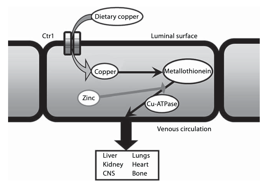
Figure : Putative copper absorption pathway. Abbreviations: Cu-ATPase = copper-transporting ATPase; CNS = central nervous system. From Choi and colleagues (Choi and Strum 2010).
Metallothionein has a higher affinity for zinc than for copper, and zinc induces further synthesis of metallothionein. Consequently, excessive zinc can effectively interfere with copper absorption at step 2 in the above sequence. As a result, copper that was brought into the enterocyte in step 1 will remain sequestered in the cell (rather than being transported into venous circulation), and will then be lost during normal epithelial cell turnover (Osadchyi et al. 2025).
Copper serves several metabolic roles, including that it functions as a cofactor in several oxidative enzymes such as cytochrome C oxidase and superoxide dismutase (Choi and Strum 2010). Copper is also involved in hematopoiesis, mitochondrial respiration and immune function (Osadchyi et al. 2025).
Neurons in the posterolateral columns are especially vulnerable to copper deficiency (Choi and Strum 2010), and this is thought to be the main mechanism underlying the corresponding myeloneuropathy.
Clinical presentation
Clinical manifestations of copper deficiency generally develop insidiously. Rounis and colleagues (Rounis et al. 2010) report an interesting exception in which a patient who had probably been gradually accumulating a copper deficiency from gastric bypass surgery and was not yet symptomatic, but became acutely symptomatic after hemodialysis.
A range of symptoms has been reported in copper deficiency, including:
- Headache (Overholser et al. 2026).
- Tinnitus (Overholser et al. 2026).
- Visual abnormalities such as blurry vision or diplopia (Rounis et al. 2010), or frank visual loss from optic neuropathy (Yarandi et al. 2014).
- Myeloneuropathic symptoms, such as:
- Paresthesias/dysesthesias, usually in the lower extremities (Choi and Strum 2010), but sometimes involving the face, tongue, neck, arms, hands or abdomen (Zara et al. 2009).
- Gait abnormalities (Overholser et al. 2026), including spastic gait (Choi and Strum 2010) and sensory ataxia (Kumar 2006).
- Vibratory sensation is reduced or absent (Choi and Strum 2010; Zara et al. 2009)
- Pinprick sensation is reduced (Choi and Strum 2010).
Copper deficiency can cause a number of significant (non-otovestibular) problems, particularly hematologic abnormalities such as anemia (microcytic, macrocytic or normocytic), leukopenia, neutropenia and thrombocytopenia (Gabreyes et al. 2013).
Clinically, the myelopathic features of copper deficiency can closely resemble subacute combined degeneration from vitamin B12 deficiency (Choi and Strum 2010; Kumar 2006).
Gabreyes and colleagues (Gabreyes et al. 2013) report that the median time from the onset of symptoms to diagnosis of zinc-induced copper deficiency was 12 months.
Physical examination
Common physical examination findings in copper deficiency include weakness, somatosensory deficits, ankle clonus, ataxic gait, and poor performance on Romberg test. Ankle reflexes are generally reduced; hypo- or hyper-reflexia can be found in other areas (Kumar et al. 2004). Rounis and colleagues (Rounis et al. 2010) report the cerebellar finding of upper extremity past-pointing.
Ocular motor examination
Rounis and colleagues (Rounis et al. 2010) report an unusual case of acute-onset copper deficiency in which physical examination showed, “bilateral horizontal nystagmus, which was more prominent and sustained on gaze to the right, as well as square wave eye jerks in the primary position of gaze.”
Testing: auditory
As of this writing there were no published reports of auditory tests in patients with copper deficiency.
Testing: vestibular
As of this writing there were no published reports of vestibular tests in patients with copper deficiency.
Testing: other
Nerve conduction velocities have been reported to show sensorimotor axonal polyneuropathy (Kumar et al. 2004). In particular, Goodman and colleagues (Goodman et al. 2006) report on a case of hypocupremic myeloneuropathy in which tibial and median somatosensory evoked potentials showed a combination of central and peripheral slowing in upper and lower limbs. Kumar and colleagues reported similar findings in 2 out of 3 patients (Kumar et al. 2004). Zara and colleagues (Zara et al. 2009) reported a case of hypocupremic myeloneuropathy in which electrodiagnostic studies showed progressive sensory action potential amplitude reduction in the median and ulnar nerves with normal sensory and motor conduction velocity.
Laboratory testing shows low serum levels of copper, low ceruloplasmin, and low urinary 24-hour copper excretion, and anemia (Kumar et al. 2004; Overholser et al. 2026).
Imaging
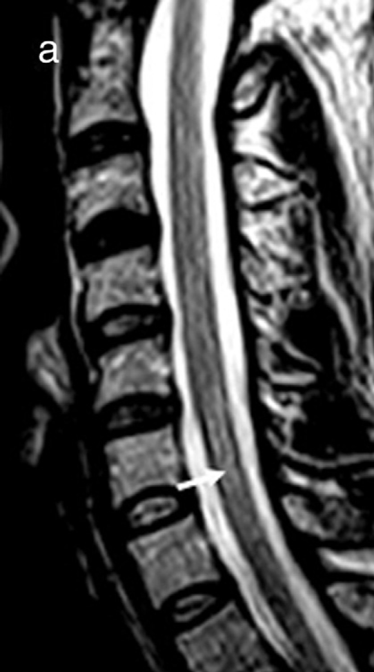
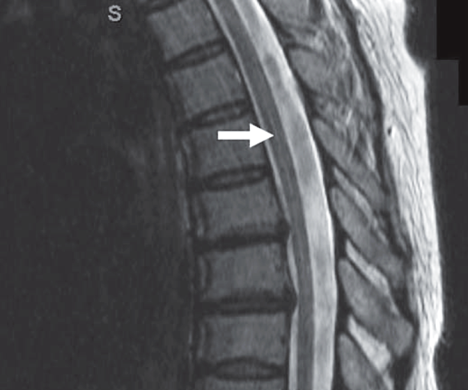
Spinal MRI in patients with hypocupremic myeloneuropathy may show T2 hyperintensity in the central cord that is longitudinal, extending over several levels in the cervical spinal cord (Gabreyes et al. 2013; Grossman and Ruiz 2021; Khaleeli et al. 2010a, b; Zara et al. 2009) or thoracic spinal cord (Choi and Strum 2010). Subacute combined degeneration from vitamin B12 deficiency can also have this appearance (Gabreyes et al. 2013).
Several cases have been reported (Goodman et al. 2006; Zara et al. 2009) in which copper supplementation brought about improvement in the signal abnormalities in the dorsal columns in the cervical spinal cord in patients with hypocupremic myeloneuropathy.
Gabreyes and colleagues (Gabreyes et al. 2013) reported that out of ten patients with copper deficiency, five (50%) exhibited multifocal T2 hyperintensities in the subcortical white matter, and 2 (20%) showed atrophy of the cerebrum and cerebellum.
The Figures below demonstrate relevant MRI abnormalities in the cervical and thoracic spinal cord in hypocupremic myeloneuropathy.
|
Figure : MRI of the cervical spine demonstrating T2 hyperintensity involving the dorsal columns (arrow). From Zara and colleagues (Zara et al. 2009) |
Figure : MRI of the thoracic spine demonstrating a faintly increased focus of T2 signal intensity (arrow) within the spinal cord in a patient with hypocupremic myeloneuropathy. From Choi and colleagues (Choi and Strum 2010). |
Histopathology
Zara and colleagues (Zara et al. 2009) report a case of hypocupremic myeloneuropathy in which a biopsy of the sural nerve showed mild axonal degeneration.
Differential diagnosis
Clinically, the myelopathic features of copper deficiency can closely resemble subacute combined degeneration from vitamin B12 deficiency (Choi and Strum 2010). The differential diagnosis includes other myelopathies and myeloneuropathies (e.g., hypovitaminosis E, folate deficiency, nitrous oxide toxicity, HIV-associated vacuolar myelopathy and neuropathy, HTLV-1 associated myelopathy, Sjögren syndrome, sarcoidosis, cervical spondylosis).
Management
Initial treatment may involve intravenous cupric sulfate (Kumar et al. 2004) or cupric chlorate (Zara et al. 2009), transitioning later to oral supplementation with cupric sulfate (Zara et al. 2009) or copper gluconate (Overholser et al. 2026).
If there is suspicion that the copper deficiency is due to zinc intake, then it is sensible to reduce dietary zinc.
Prognosis
Literature generally reports that despite appropriate treatment with copper supplementation, the neurological outcomes are poor. Most studies report that even with treatment, the neurological deficits in copper deficiency sometimes worsen (Gabreyes et al. 2013), are often irreversible (Gabreyes et al. 2013), or show only modest improvement (Jaiser and Winston 2010). Early recognition and treatment lead to more favorable outcomes (Osadchyi et al. 2025).
Gabreyes and colleagues (Gabreyes et al. 2013) reported that out of 12 patients three (25%) exhibited partial improvement in sensory and motor deficits, five (42%) showed no change, and four (33%) further deteriorated and became wheelchair-bound.
Goodman and colleagues (Goodman et al. 2006) reported a case of hypocupremic myeloneuropathy in which supplementation of copper brought about some improvement in strength, but no changes in the sensory deficits.
Overholser and colleagues (Overholser et al. 2026) describe a case in which copper supplementation brought about improvement in neuropathy.
Kumar and colleagues (Kumar et al. 2004) describe three cases, one of which reported some improvement in gait with copper supplementation. Rounis and colleagues (Rounis et al. 2010) also report a case in which there was modest improvement in gait.
It has been reported that hypocupremia-induced tinnitus resolved with copper supplementation (Overholser et al. 2026).
The generally poor neurologic outcomes contrast sharply with the hematologic parameters, that usually normalize with copper supplementation (Gabreyes et al. 2013).
References
Carroll LS, Abdul-Rahim AH, Murray R (2017) Zinc containing dental fixative causing copper deficiency myelopathy. BMJ Case Rep 2017. doi: 10.1136/bcr-2017-219802
Choi EH, Strum W (2010) Hypocupremia-related myeloneuropathy following gastrojejunal bypass surgery. Ann Nutr Metab 57: 190-2. doi: 10.1159/000321519
Gabreyes AA, Abbasi HN, Forbes KP, McQuaker G, Duncan A, Morrison I (2013) Hypocupremia associated cytopenia and myelopathy: a national retrospective review. Eur J Haematol 90: 1-9. doi: 10.1111/ejh.12020
Goodman BP, Chong BW, Patel AC, Fletcher GP, Smith BE (2006) Copper deficiency myeloneuropathy resembling B12 deficiency: partial resolution of MR imaging findings with copper supplementation. AJNR Am J Neuroradiol 27: 2112-4.
Grossman JT, Ruiz S (2021) Copper Deficiency Myeloneuropathy in Autoimmune Disease. Cureus 13: e16591. doi: 10.7759/cureus.16591
Jaiser SR, Winston GP (2010) Copper deficiency myelopathy. J Neurol 257: 869-81. doi: 10.1007/s00415-010-5511-x
Khaleeli Z, Healy DG, Briddon A, Lunn MP, Reilly MM, Land J, Giovannoni G (2010a) Copper deficiency as a treatable cause of poor balance. BMJ 340: c508. doi: 10.1136/bmj.c508
Khaleeli Z, Healy DG, Briddon A, Lunn MP, Reilly MM, Land J, Giovannoni G (2010b) Kupfermangel als behandelbare Ursache von Gleichgewichtsstörungen [Copper deficiency as a treatable cause of poor balance]. Praxis (Bern 1994) 99: 1153-6. doi: 10.1024/1661-8157/a000258
Kumar N (2006) Copper deficiency myelopathy (human swayback). Mayo Clin Proc 81: 1371-84. doi: 10.4065/81.10.1371
Kumar N, Crum B, Petersen RC, Vernino SA, Ahlskog JE (2004) Copper deficiency myelopathy. Arch Neurol 61: 762-6. doi: 10.1001/archneur.61.5.762
Osadchyi V, Van Antwerp SN, Vredenburgh J (2025) Zinc-Induced Copper Deficiency Myeloneuropathy Masquerading as Paraneoplastic Syndrome: A Case Report. Cureus 17: e82995. doi: 10.7759/cureus.82995
Overholser A, Landis E, Szymanski K (2026) Copper deficiency myelopathy and sensory ataxia in a 20-year-old female. JAAPA 39: e2-e4. doi: 10.1097/01.JAA.0000000000000321
Rounis E, Laing CM, Davenport A (2010) Acute neurological presentation due to copper deficiency in a hemodialysis patient following gastric bypass surgery. Clin Nephrol 74: 389-92.
Rowin J, Lewis SL (2005) Copper deficiency myeloneuropathy and pancytopenia secondary to overuse of zinc supplementation. J Neurol Neurosurg Psychiatry 76: 750-1. doi: 10.1136/jnnp.2004.046987
Yarandi SS, Griffith DP, Sharma R, Mohan A, Zhao VM, Ziegler TR (2014) Optic neuropathy, myelopathy, anemia, and neutropenia caused by acquired copper deficiency after gastric bypass surgery. J Clin Gastroenterol 48: 862-5. doi: 10.1097/MCG.0000000000000092
Zara G, Grassivaro F, Brocadello F, Manara R, Pesenti FF (2009) Case of sensory ataxic ganglionopathy-myelopathy in copper deficiency. J Neurol Sci 277: 184-6. doi: 10.1016/j.jns.2008.10.017
