By Marcello Cherchi, MD PhD
For patients
Orthostatic hypotension is a condition in which going from a lying down position to a seated or standing position causes a large drop in blood pressure. That drop in blood pressure can cause symptoms such as dizziness, visual changes, lightheadedness, and fainting (loss of consciousness). If your doctor suspects a diagnosis of orthostatic hypotension, then they may check several tests. Some patients manage this problem without medications, while others use medications.
For clinicians
Overview
Orthostatic hypotension is a type of orthostatic intolerance in which going from a supine/prone position to an upright (standing more than sitting) position provokes an inappropriately large drop in blood pressure, which can manifest with symptoms of disequilibrium, visual changes, presyncope and syncope. Orthostatic hypotension is more common with advancing age. Some investigators classify orthostatic hypotension into two types, namely “neurogenic” (e.g., as can occur in some parkinsonian disorders, familial dysautonomia, autoimmune autonomic ganglionopathy) and “non-neurogenic” (e.g., as can occur with some medications, diabetes, various cardiovascular and renal diseases, some endocrinological states, volume depletion, spinal cord lesions, and others). Orthostatic hypotension can often be identified on bedside examination, but tilt table testing is more sensitive. Cerebral blood flow is imperfectly correlated with peripherally measured blood pressure, so if the clinical index of suspicion remains high (despite a negative tilt table test), there may be some rationale for empiric treatment. Management of orthostatic hypotension depends on the underlying cause (if discernible); it often starts with non-pharmacologic strategies before progressing to pharmacologic treatment.
Introduction
In simple terms, orthostatic hypotension is a type of orthostatic intolerance in which going from a supine/prone position to an upright (standing more than sitting) position provokes an inappropriately large drop in blood pressure, which can manifest with symptoms of disequilibrium, visual changes, presyncope and syncope.
Epidemiology
Literature describes the prevalence of orthostatic hypotension as being age-dependent, with figures such as:
- 5% of people under 50 years of age (Ricci et al. 2015).
- 16% of people over 65 years of age (Shibao and Biaggioni 2020).
- 30% of people over 70 years of age (Ricci et al. 2015).
Genetics
Orthostatic hypotension in itself is not known to have a specific genetic mechanism. However, there are some genetically transmitted disorders (such as dopamine-beta-hydroxylase deficiency, OMIM 609312) whose manifestations include orthostatic hypotension.
Pathophysiological mechanism of disease
Ricci and colleagues (Ricci et al. 2015) comment that:
“Orthostatic stress is a common daily challenge for humans when posture changes from lying to standing or during prolonged quiet standing. Almost immediately, with the transition from the supine (recumbent) to the upright (erect) position, a gravitational shift of nearly 500 mL of blood away from the chest to the distensible venous capacitance system below the diaphragm (venous pooling) occurs. This results in a rapid decrease in central blood volume and a subsequent reduction of ventricular preload, stroke volume, and mean BP” (Ricci et al. 2015).
A schematic of these hemodynamic changes is depicted in Figure 1 below from Moloney and colleagues (Moloney et al. 2026).
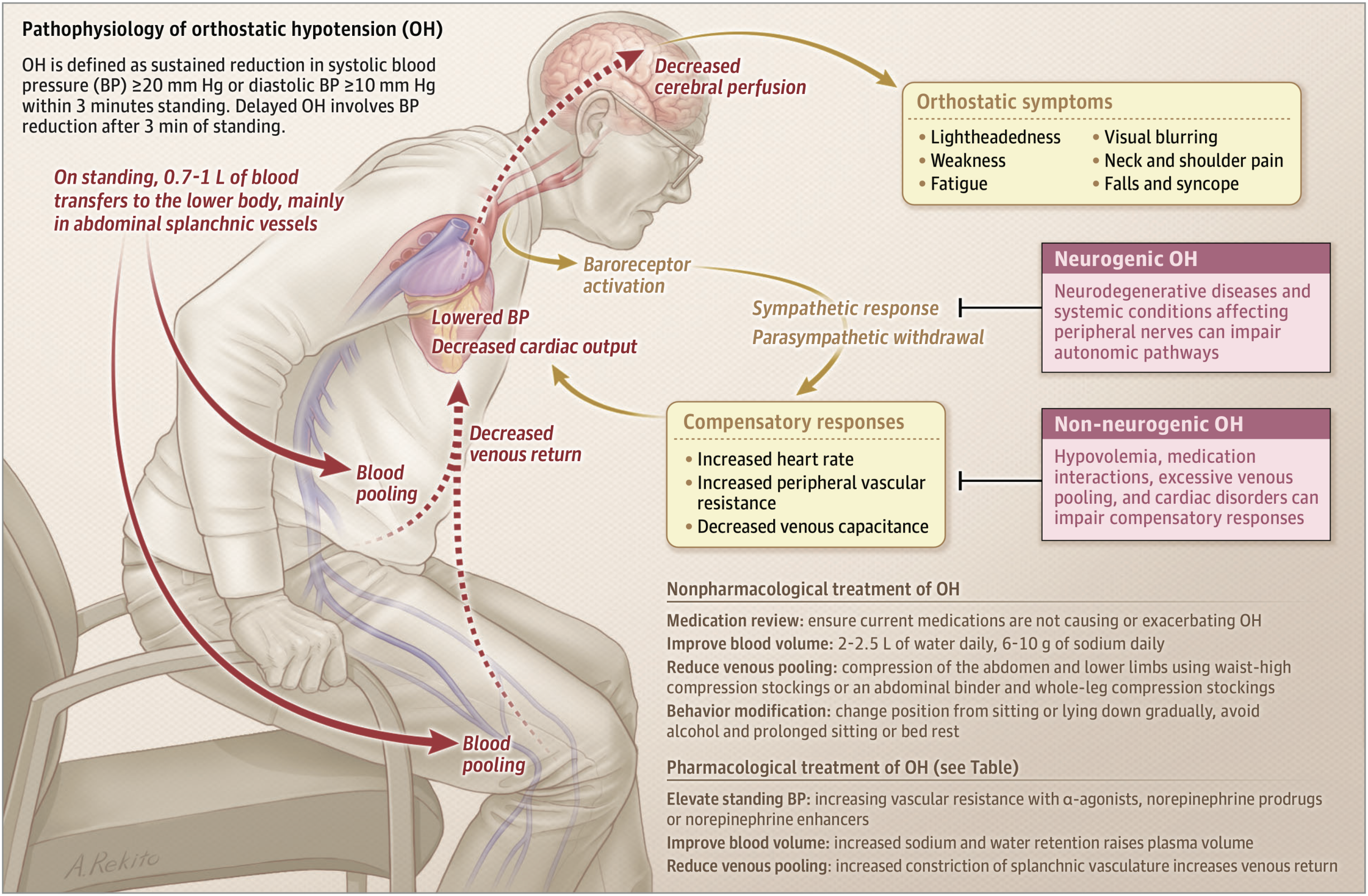
Figure : Pathophysiology of orthostatic hypotension. From Moloney et al (Moloney et al. 2026).
The autonomic nervous system maintains blood pressure based on a variety of inputs, predominantly afferent signaling from baroreceptors located in the carotid sinus and aortic arch. That information is processed in the vasomotor center of the medulla, which then sends sympathetic and parasympathetic efferent projections to the heart and vascular system, as well as projections to the hypothalamus (to activate endocrine mechanisms when necessary).
A schematic of this circuitry is depicted in Figure 2 below from Ricci and colleagues (Ricci et al. 2015).
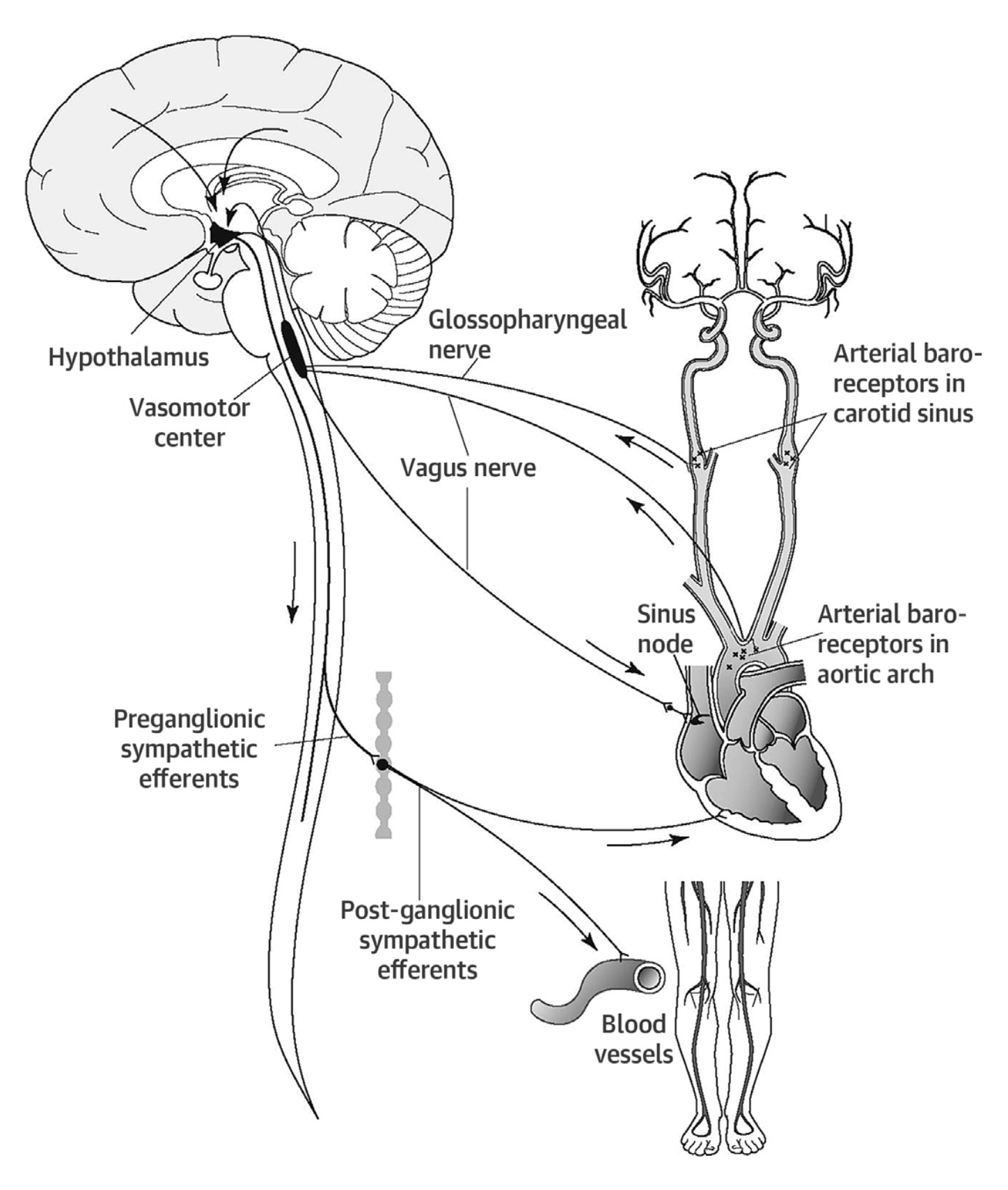
Figure : Schematic of circuitry involved in maintenance of blood pressure and cardiac pulse. The afferent pathway transfers information from arterial baroreceptors in the carotid sinus and aortic arch. This information reaches the vasomotor center in the medulla oblongata. The efferent pathway regulates two basic cardiovascular responses: heart rate and vascular tone (blood pressure). The hypothalamus can then activate vasopressin release as an additional regulatory mechanism. Higher brain functions can modulate autonomic cardiovascular responses. From Ricci et al (Ricci et al. 2015).
Disease processes that interfere with the afferent or efferent pathways (or both) of this circuit have the potential to impair regulation of blood pressure and heart rate.
Ricci and colleagues (Ricci et al. 2015) broadly classify orthostatic hypotension into primary and secondary forms:
- Primary orthostatic hypotension (sometimes also called “neurogenic orthostatic hypotension”):
- Parkinson’s disease
- Multiple system atrophy
- Pure autonomic failure
- Lewy body dementia
- Autoimmune autonomic ganglionopathy
- Rare hereditary disorders (familial dysautonomia, dopamine beta-hydroxylase deficiency)
- Idiopathic (etiology unknown)
- Secondary orthostatic hypotension (sometimes also called “non-neurogenic orthostatic hypotension”):
- Iatrogenic (e.g., pharmacologic)
- Diabetes mellitus
- Cardiovascular diseases (sick sinus syndrome, atrioventricular block, heart failure, aortic stenosis, pulmonary hypertension, essential hypertension)
- Renal failure
- Autoimmune diseases
- Volume depletion
- Venous pooling
- Alcoholic polyneuropathy
- Endocrine disorders (adrenal insufficiency, thyroid diseases, diabetes insipidus)
- Amyloidosis
- Multiple myeloma
- Paraneoplastic syndromes
- Cerebrovascular disease
- Multiple sclerosis
- Spinal cord diseases
Theoretical considerations
Most studies on orthostatic intolerance (including orthostatic hypotension) tacitly assume that heart rate and peripheral measurement of blood pressure (using a regular sphygmomanometer) serve as an adequate proxy for cerebral blood flow, but this assumption may be mistaken. Van Campen and colleagues (van Campen et al. 2020) studied healthy controls and chronic fatigue syndrome (myalgic encephalomyelitis) patients during tilt table testing using carotid Doppler flow studies, and reported that cerebral blood flow (as measured by carotid Doppler) could still be impaired even when assessment by traditional tilt table parameters (for orthostatic hypotension and postural orthostatic tachycardia) were normal.
Clinical presentation
Orthostatic hypotension “may provoke signs and symptoms of cerebral hypoperfusion, including nausea, fatigue, lightheadedness… visual blurring, and eventually syncope” (Ricci et al. 2015).
Physical examination
Typical consensus criteria for orthostatic hypotension are that going from a supine to a standing position elicits a drop in systolic blood pressure of ≥20 mm Hg, or a drop in diastolic blood pressure of ≥10 mm Hg, or both, within 3 minutes of the positional change (Kaufmann 1996; Schatz et al. 1996a, b, c). The stated time limitation (“within 3 minutes”) essentially pertains to cases that clinicians can detect at the bedside; in practice this criterion will miss cases in which the drop in blood pressure is delayed (Gibbons and Freeman 2020), which may only be detected on tilt table testing (TTT).
Ocular motor examination
Ocular motor examination generally does not provide evidence to support or refute a diagnosis of orthostatic hypotension.
Testing: auditory
Auditory testing generally does not provide evidence to support or refute a diagnosis of orthostatic hypotension.
Testing: vestibular
Choi and colleagues (Choi et al. 2015) studied 33 patients with orthostatic hypotension with infrared video oculography (VOG) during the Schellong test (supine to active standing, described by Dr. Fritz Schellong (Schellong 1938)) and squatting-to-standing test, and reported that 10 (30%) of these patients developed nystagmus upon standing; specifically, 4 exhibited nystagmus that was down beat, while 5 exhibited nystagmus that was down beat plus horizontal (and some had a torsional component).
This is an interesting observation, but not a common application of infrared video oculography (VOG) in routine clinical practice.
Testing: other
Tilt table testing (TTT) can be helpful in diagnosing and characterizing orthostatic hypotension. It is especially helpful in cases of delayed orthostatic hypotension, in which the requisite drop in blood pressure does not happen until after the usual “within 3 minutes” criterion.
Examples of regular orthostatic hypotension and delayed orthostatic hypotension (culminating in syncope) are shown in the Figure 3 below from Ricci and colleagues (Ricci et al. 2015).
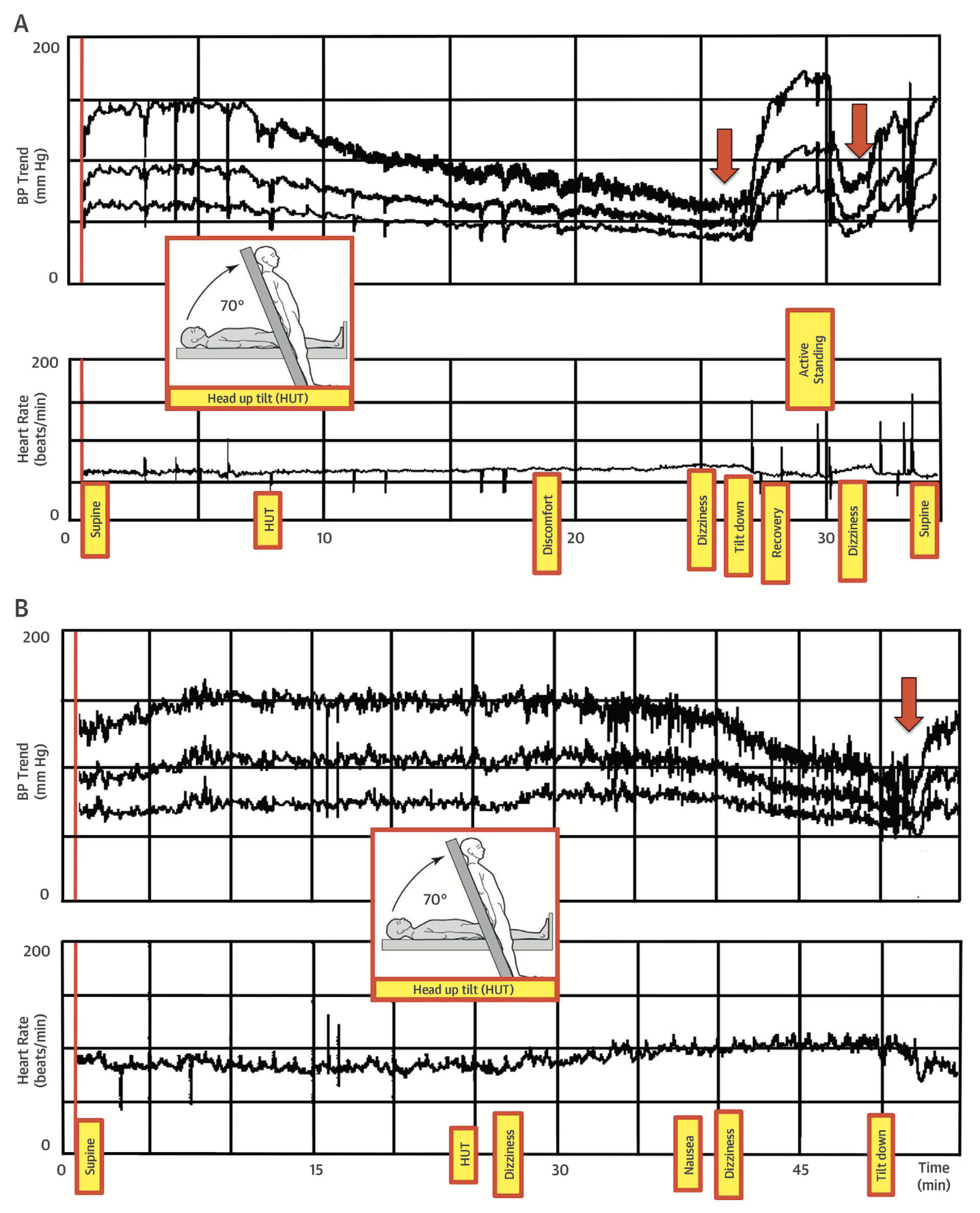
Figure : Examples of tilt table test results in patients with immediate and delayed orthostatic hypotension. The upper tracing in each panel shows a beat-to-beat blood pressure (BP) measurement and consecutive test stages. The lower tracing in each panel shows the heart rate. Red arrows mark the moment when syncope occurs. Panel A shows the results of a 52-year-old woman with classic (immediate) orthostatic hypotension and reflex syncope during passive head-up tilt (HUT). Panel B shows the results of a 74-year-old woman with delayed orthostatic hypotension and syncope during passive HUT. From Ricci et al (Ricci et al. 2015).
A home device for continuous blood pressure monitoring (usually dispensed by cardiology) may also be helpful, particularly if the patient can maintain a corresponding symptom log.
Imaging
Imaging of the neuraxis generally does not provide evidence to support or refute a diagnosis of orthostatic hypotension. Specific exceptions to this include imaging for spinal cord injuries, some of which can interfere with autonomic function.
Histopathology
Histopathological studies generally do not provide evidence to support or refute a diagnosis of orthostatic hypotension. Specific exceptions include post-mortem histopathology in neurodegenerative disorders (such as multiple system atrophy) that are associated with autonomic dysfunction.
Differential diagnosis
The differential diagnosis of orthostatic hypotension includes the other categories of orthostatic intolerance (orthostatic hypertension, postural orthostatic tachycardia syndrome).
Robertson and Robertson (Robertson and Robertson 1994) suggest the following extensive differential diagnosis of orthostatic hypotension:
- Autonomic disorders
- Bradbury-Eggleston syndrome (pure autonomic failure)
- Shy-Drager syndrome (multiple system atrophy)
- Autonomic failure with Parkinson’s disease
- Riley-Day syndrome (familial dysautonomia)
- Dopamine-beta-hydroxylase deficiency
- Baroreceptor failure
- Acute pandysautonomia
- Diabetes mellitus
- Amyloidosis
- Porphyria
- Paraneoplastic autonomic neuropathy
- Acute inflammatory neuropathy
- Hereditary sensory neuropathy
- Bradbury-Eggleston syndrome (pure autonomic failure)
- Hypovolemic disorders
- Hemorrhage or plasma loss
- Overdiuresis
- Overdialysis
- Idiopathic hypovolemia
- Hemorrhage or plasma loss
- Endocrinologic disorders
- Addison’s disease
- Hypoaldosteronism
- Pheochromocytoma
- Renovascular hypertension
- Addison’s disease
- Vascular insufficiency
- Varicose veins
- Absent venous valves
- Arteriovenous malformations
- Varicose veins
- Vasodilator excess
- Mastocytosis
- Hyperbradykininism (bradykinin)
- Carcinoid (bradykinin)
- Hypermagnesemia
- Mastocytosis
- Paroxysmal autonomic syncope
- Glossopharyngeal syncope
- Micturation syncope
- Carotid sinus syndrome
- Swallow syncope
- Cough syncope
- Bezold-Jarisch reflex activation
- Glossopharyngeal syncope
- Miscellaneous
- Drugs and toxins
- Stokes-Adams attacks
- Mitral valve prolapse syndrome
- Gastrectomy
- Hypokinesia, weightlessness, bed rest
- Drugs and toxins
Management: non-pharmacologic strategies
Several non-pharmacologic strategies have been studied for managing orthostatic hypotension.
Review medications with the prescribing physicians
Review medications with the physicians who prescribed them to ascertain whether any may be contributing to (or causing) orthostatic hypotension. If so, then discuss whether it is possible to eliminate, reduce, or substitute any of those medications. Medications intended to lower blood pressure can cause or worsen orthostatic intolerance. However, many other medications have the unintended effect of lowering blood pressure, or causing blood pressure to fluctuate. The patient should be reminded not simply to stop medications without first consulting the physicians currently prescribing them.
Increase hydration
Increased hydration has been shown to be an effective strategy for patients (Jordan et al. 2000; May and Jordan 2011; Newton and Frith 2018). There are no exact criteria on how much of an increase is appropriate; a goal of 2 to 2.5 liters per day is frequently cited (Moloney et al. 2026). It is preferable, when possible, to drink water, or water containing electrolytes (such as Gatorade® or Pedialyte®). The strategy of increasing fluid intake may not be appropriate in all patients; for instance, patients with kidney failure or heart failure may be advised by their doctor not to increase fluid intake.
Increase sodium intake
Eating sodium (usually in the form of table salt) helps the body to maintain more effectively any fluids already ingested, and thus can help raise and stabilize blood pressure, which can reduce the risk of orthostatic hypotension. This strategy may not be appropriate for all people, since some patients have other medical conditions for which dietary sodium intake should be decreased; for instance, many patients with cardiac disease are specifically instructed to reduce salt in their diet.
Wear compression stockings
Compression stockings (sometimes also called Jobst stockings) are basically tights. By squeezing the legs, pelvis and lower abdomen, they reduce the amount of blood that pools there, and have been shown to decrease the risk of orthostatic hypotension (Denq et al. 1997; Podoleanu et al. 2006). Ideally the stockings should come up to the waist, because this prevents pooling of blood not only in the legs but also in the pelvic venous plexus. Stockings that only come up to the knee or to the hip are less effective. Compression stockings can be obtained in medical supply stores. They do not require a prescription in the United States. There are different degrees of “tightness.” The patient should choose a grade of tightness that they can tolerate. If the stockings are too loose, then they will be ineffective or less effective.
Wear an abdominal binder
An abdominal binder resembles a girdle. By modestly compressing the abdomen, it promotes the return of blood from the abdominal venous plexus up into the thorax, and can benefit patients with orthostatic hypotension (Figueroa et al. 2015; Smit et al. 2004). Abdominal binders are available at medical supply stores. They come in different sizes and degrees of tightness, and do not require a prescription in the United States.
Adjust the bed so that the head is higher than the pelvis, and the pelvis is higher than the feet
Adjusting the bed so that the head is higher than the pelvis and the pelvis is higher than the feet is called the “reverse Trendelenburg position.” This strategy maintains modest blood pooling in the lower extremities at night and thereby keeps the renin-angiotensin-aldosterone axis slightly activated (van der Stam et al. 2023), such that when the patient arises in the morning, they will be less likely to suffer a large drop in blood pressure. Note that it is not adequate simply to sleep with a few extra pillows beneath the head; rather, the whole bed must be inclined. Usually this is most easily accomplished by putting 1 or 2 bricks beneath each post at the head of the bed. An angle of at least 10˚ is appropriate (Moloney et al. 2026).
Avoid eating large meals
After ingesting a large meal, the body purposefully shunts up to 20% of the blood to the gastrointestinal tract to aid in digestion (Victor and Ropper 2001). This means that less blood is available for other tasks, such as circulating to the brain. When feasible, it is preferable to eat a greater number of small meals rather than a smaller number of large meals. This does not mean that a patient needs to reduce their total daily food intake; rather, it means that the total food intake should be distributed more evenly throughout the day. In other words, it is better to “graze” rather than “feast.”
Avoid hot environments
When a person feels warm, or when perspiring, the body purposefully shunts blood to circulate just beneath the skin in an attempt to dissipate heat (Okamoto et al. 2021), one consequence of which is that less blood is available for other tasks, such as circulating to the brain, which can cause or exacerbate orthostatic hypotension. When possible, avoid hot environments.
Avoid straining at stool
The act of straining — such as when moving one’s bowels or when lifting heavy objects — is called the Valsalva maneuver. At the beginning of the Valsalva maneuver there is a brief increase in blood pressure, followed by a decrease in blood pressure that persists both during the remainder maneuver itself, and after “releasing” from the straining (Looga 2005). If constipation is causing a patient to strain during bowel movements, then it is sensible to try to alleviate the constipation with increased hydration, increased dietary fiber, prunes, or laxatives.
Get out of bed gradually
Going from a supine or prone position to a standing position poses a risk of orthostatic hypotension, and this risk is higher after prolonged bedrest (Kamiya et al. 2003). Most people with orthostatic hypotension instinctively figure out that they need to get out of bed slowly — basically, transition from a supine to a standing position in stages with pauses at each stage. For example, after waking up, first sit at the side of the bed for a few minutes before actually standing up to walk. This strategy reduces the risk of orthostatic hypotension (de Bruine et al. 2017).
Orthostatic training
Orthostatic training consists of several relatively simple exercises that a patient can learn from a physical therapist familiar with the problem of orthostatic hypotension.
The main exercise is simple and can be done at home. It consists simply of standing up against a wall for 30 minutes. Initially patients may find they are unable to maintain this posture for a full 30 minutes, but over time they increase their tolerance and usually are able to achieve this target. One study of an orthostatic training program followed 24 patients who actually suffered frequent fainting due to their orthostatic hypotension (Abe et al. 2002). The intention was for patients to stand against a wall without moving for 30 minutes twice per day. However, most patients only ended up doing the exercise once per day. Nevertheless, after 5 to 18 months, 22 patients (92%) no longer suffered syncope. Most other studies have found similar results (Abe et al. 2003a; Abe et al. 2003b; Ector et al. 1998; Kinay et al. 2004; Reybrouck and Ector 2006; Reybrouck et al. 2000). However, a minority of studies found no significant difference between those patients who performed the exercises and those who did not (Duygu et al. 2008; Foglia-Manzillo et al. 2004; On et al. 2007).
For patients who have failed other therapies, we usually recommend trying orthostatic training since there is no significant medical risk. The more frequently a patient performs the exercises, and the greater the length of time (i.e., preferably months rather than simply weeks), the more likely it is that they will benefit from them.
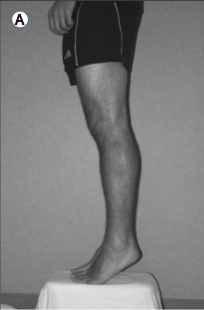
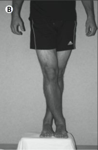
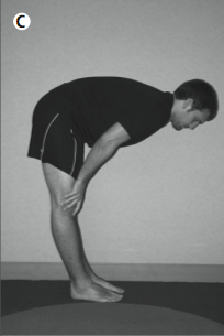
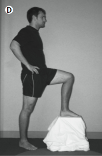
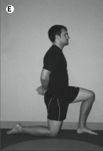
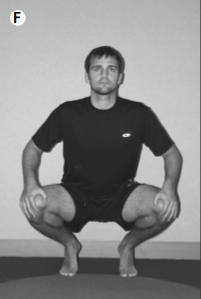
Even when the above-mentioned exercise is helpful, some patients may still experience occasional lightheadedness when upright. If a patient has this sensation and fears that they might faint, then there are several “counter-maneuvers” that reduce its likelihood. The maneuvers include toe raising, leg crossing, thigh contraction, and bending at the waist, all of which reduce venous capacity and increase total peripheral resistance (Low and Singer 2008). Illustrations of these maneuvers are given below.
| 
| 
| 
| 
| 
|
Toe raise | Leg cross | Forward lean | Step up | Genuflection | Squat |
Management: pharmacologic strategies
Several pharmacologic strategies have been studied for managing orthostatic hypotension. Two drugs (midodrine and droxidopa) have received FDA-approval for management of orthostatic hypotension, while the others (fludrocortisone, atomoxetine and pyridostigmine) have been used off-label for this purpose.
Midodrine is an alpha-1 adrenergic receptor agonist whose sympathomimetic effects are superior to placebo in managing orthostatic hypotension (Fouad-Tarazi et al. 1995; Jankovic et al. 1993; Low et al. 1997; Wright et al. 1998), though a systematic review and meta-analysis judged the overall evidence to be of limited quality (Parsaik et al. 2013). Common adverse effects of midodrine include supine hypertension and urinary retention. In the US, as of this writing, midodrine was FDA-approved for management of neurogenic and non-neurogenic orthostatic hypotension.
Droxidopa is a norepinephrine precursor. Studies have shown it to improve orthostatic blood pressure and symptoms of orthostatic hypotension (Biaggioni et al. 2017). In the US, as of this writing, droxidopa was FDA-approved for management of neurogenic orthostatic hypotension. Compared to midodrine, droxidopa may be less efficacious, but also less likely to cause supine hypertension (Chen et al. 2018).
Fludrocortisone has glucocorticoid and mineralocorticoid effects, and has been used off-label in the management of orthostatic hypotension for several decades. As of this writing, fludrocortisone was not FDA-approved for management of orthostatic hypotension. A Cochrane review concluded that the evidence supporting the efficacy of fludrocortisone was weak (Veazie et al. 2021). Potential adverse effects of fludrocortisone include supine hypertension, hypokalemia, and (in patients with a history of cardiac failure) decompensation of (previously compensated) heart failure.
Atomoxetine was originally developed for the treatment of attention deficit disorder. Atomoxetine inhibits the activity of a presynaptic norepinephrine transporter, thereby decreasing norepinephrine reuptake in postganglionic sympathetic neurons (Shibao et al. 2007). As of this writing, atomoxetine was not FDA-approved for management of orthostatic hypotension. Potential adverse effects of atomoxetine include supine hypertension, tachycardia, dry mouth and insomnia.
Pyridostigmine was originally developed for the treatment of myasthenia gravis. Pyridostigmine inhibits acetylcholinesterase, thereby facilitating cholinergic transmission in autonomic ganglia, thereby augmenting sympathetic tone. These properties make its use logical in neurogenic orthostatic hypotension (Singer et al. 2003). A randomized, double-blind, placebo controlled crossover trial of pyridostigmine in the management of neurogenic orthostatic hypotension in 58 patients showed pyridostigmine’s superiority (over placebo) in improving orthostatic hypotension and its symptoms, without causing supine hypertension. As of this writing, pyridostigmine was not FDA-approved for management of orthostatic hypotension. Potential adverse effects include abdominal cramping, diarrhea, urinary incontinence, sialorrhea and diaphoresis.
Prognosis
Most literature regarding “prognosis” of orthostatic hypotension pertains not to the success or failure of managing orthostatic hypotension itself, but rather the prognostic value that orthostatic hypotension has pertaining to other problems such as various cardiovascular morbidities or all-cause mortality (Ricci et al. 2015).
References
Abe H, Kohshi K, Nakashima Y (2003a) Effects of orthostatic self-training on head-up tilt testing and autonomic balance in patients with neurocardiogenic syncope. J Cardiovasc Pharmacol 41 Suppl 1: S73-6.
Abe H, Kondo S, Kohshi K, Nakashima Y (2002) Usefulness of orthostatic self-training for the prevention of neurocardiogenic syncope. Pacing Clin Electrophysiol 25: 1454-8.
Abe H, Sumiyoshi M, Kohshi K, Nakashima Y (2003b) Effects of orthostatic self-training on head-up tilt testing for the prevention of tilt-induced neurocardiogenic syncope: comparison of pharmacological therapy. Clin Exp Hypertens 25: 191-8.
Biaggioni I, Arthur Hewitt L, Rowse GJ, Kaufmann H (2017) Integrated analysis of droxidopa trials for neurogenic orthostatic hypotension. BMC Neurol 17: 90. doi: 10.1186/s12883-017-0867-5
Chen JJ, Han Y, Tang J, Portillo I, Hauser RA, Dashtipour K (2018) Standing and Supine Blood Pressure Outcomes Associated With Droxidopa and Midodrine in Patients With Neurogenic Orthostatic Hypotension: A Bayesian Meta-analysis and Mixed Treatment Comparison of Randomized Trials. Ann Pharmacother 52: 1182-1194. doi: 10.1177/1060028018786954
Choi JH, Seo JD, Kim MJ, Choi BY, Choi YR, Cho BM, Kim JS, Choi KD (2015) Vertigo and nystagmus in orthostatic hypotension. Eur J Neurol 22: 648-55. doi: 10.1111/ene.12622
de Bruine ES, Reijnierse EM, Trappenburg MC, Pasma JH, de Vries OJ, Meskers CG, Maier AB (2017) Standing Up Slowly Antagonises Initial Blood Pressure Decrease in Older Adults with Orthostatic Hypotension. Gerontology 63: 137-143. doi: 10.1159/000450642
Denq JC, Opfer-Gehrking TL, Giuliani M, Felten J, Convertino VA, Low PA, New Collective A (1997) Efficacy of compression of different capacitance beds in the amelioration of orthostatic hypotension. Clin Auton Res 7: 321-6. doi: 10.1007/BF02267725
Duygu H, Zoghi M, Turk U, Akyuz S, Ozerkan F, Akilli A, Erturk U, Onder R, Akin M (2008) The role of tilt training in preventing recurrent syncope in patients with vasovagal syncope: a prospective and randomized study. Pacing Clin Electrophysiol 31: 592-6. doi: 10.1111/j.1540-8159.2008.01046.x
Ector H, Reybrouck T, Heidbuchel H, Gewillig M, Van de Werf F (1998) Tilt training: a new treatment for recurrent neurocardiogenic syncope and severe orthostatic intolerance. Pacing Clin Electrophysiol 21: 193-6.
Figueroa JJ, Singer W, Sandroni P, Sletten DM, Gehrking TL, Gehrking JA, Low P, Basford JR (2015) Effects of patient-controlled abdominal compression on standing systolic blood pressure in adults with orthostatic hypotension. Arch Phys Med Rehabil 96: 505-10. doi: 10.1016/j.apmr.2014.10.012
Foglia-Manzillo G, Giada F, Gaggioli G, Bartoletti A, Lolli G, Dinelli M, Del Rosso A, Santarone M, Raviele A, Brignole M (2004) Efficacy of tilt training in the treatment of neurally mediated syncope. A randomized study. Europace 6: 199-204. doi: 10.1016/j.eupc.2004.01.002S1099512904000042
Fouad-Tarazi FM, Okabe M, Goren H (1995) Alpha sympathomimetic treatment of autonomic insufficiency with orthostatic hypotension. Am J Med 99: 604-10. doi: 10.1016/s0002-9343(99)80246-0
Gibbons CH, Freeman R (2020) Delayed orthostatic hypotension. Auton Neurosci 229: 102724. doi: 10.1016/j.autneu.2020.102724
Jankovic J, Gilden JL, Hiner BC, Kaufmann H, Brown DC, Coghlan CH, Rubin M, Fouad-Tarazi FM (1993) Neurogenic orthostatic hypotension: a double-blind, placebo-controlled study with midodrine. Am J Med 95: 38-48. doi: 10.1016/0002-9343(93)90230-m
Jordan J, Shannon JR, Black BK, Ali Y, Farley M, Costa F, Diedrich A, Robertson RM, Biaggioni I, Robertson D, New Collective A (2000) The pressor response to water drinking in humans : a sympathetic reflex? Circulation 101: 504-9. doi: 10.1161/01.cir.101.5.504
Kamiya A, Michikami D, Fu Q, Iwase S, Hayano J, Kawada T, Mano T, Sunagawa K (2003) Pathophysiology of orthostatic hypotension after bed rest: paradoxical sympathetic withdrawal. Am J Physiol Heart Circ Physiol 285: H1158-67. doi: 10.1152/ajpheart.00965.2002
Kaufmann H (1996) Consensus statement on the definition of orthostatic hypotension, pure autonomic failure and multiple system atrophy. Clin Auton Res 6: 125-6.
Kinay O, Yazici M, Nazli C, Acar G, Gedikli O, Altinbas A, Kahraman H, Dogan A, Ozaydin M, Tuzun N, Ergene O (2004) Tilt training for recurrent neurocardiogenic syncope: effectiveness, patient compliance, and scheduling the frequency of training sessions. Jpn Heart J 45: 833-43. doi: JST.JSTAGE/jhj/45.833 [pii]
Looga R (2005) The Valsalva manoeuvre–cardiovascular effects and performance technique: a critical review. Respir Physiol Neurobiol 147: 39-49.
Low PA, Gilden JL, Freeman R, Sheng KN, McElligott MA (1997) Efficacy of midodrine vs placebo in neurogenic orthostatic hypotension. A randomized, double-blind multicenter study. Midodrine Study Group. JAMA 277: 1046-51.
Low PA, Singer W (2008) Management of neurogenic orthostatic hypotension: an update. Lancet Neurol 7: 451-8. doi: 10.1016/S1474-4422(08)70088-7
May M, Jordan J (2011) The osmopressor response to water drinking. Am J Physiol Regul Integr Comp Physiol 300: R40-6. doi: 10.1152/ajpregu.00544.2010
Moloney D, Youssef A, Okamoto LE (2026) Management of Orthostatic Hypotension: A Review. JAMA Intern Med. doi: 10.1001/jamainternmed.2026.0284
Newton JL, Frith J (2018) The efficacy of nonpharmacologic intervention for orthostatic hypotension associated with aging. Neurology 91: e652-e656. doi: 10.1212/WNL.0000000000005994
Okamoto LE, Celedonio JE, Smith EC, Gamboa A, Shibao CA, Diedrich A, Paranjape SY, Black BK, Muldowney JAS, 3rd, Peltier AC, Habermann R, Crandall CG, Biaggioni I (2021) Local Passive Heat for the Treatment of Hypertension in Autonomic Failure. J Am Heart Assoc 10: e018979. doi: 10.1161/JAHA.120.018979
On YK, Park J, Huh J, Kim JS (2007) Is home orthostatic self-training effective in preventing neurally mediated syncope? Pacing Clin Electrophysiol 30: 638-43. doi: 10.1111/j.1540-8159.2007.00725.x
Parsaik AK, Singh B, Altayar O, Mascarenhas SS, Singh SK, Erwin PJ, Murad MH (2013) Midodrine for orthostatic hypotension: a systematic review and meta-analysis of clinical trials. J Gen Intern Med 28: 1496-503. doi: 10.1007/s11606-013-2520-3
Podoleanu C, Maggi R, Brignole M, Croci F, Incze A, Solano A, Puggioni E, Carasca E (2006) Lower limb and abdominal compression bandages prevent progressive orthostatic hypotension in elderly persons: a randomized single-blind controlled study. J Am Coll Cardiol 48: 1425-32. doi: 10.1016/j.jacc.2006.06.052
Reybrouck T, Ector H (2006) Tilt training: a new challenge in the treatment of neurally mediated syncope. Acta Cardiol 61: 183-9.
Reybrouck T, Heidbuchel H, Van de Werf F, Ector H (2000) Tilt training: a treatment for malignant and recurrent neurocardiogenic syncope. Pacing Clin Electrophysiol 23: 493-8.
Ricci F, De Caterina R, Fedorowski A (2015) Orthostatic Hypotension: Epidemiology, Prognosis, and Treatment. J Am Coll Cardiol 66: 848-860. doi: 10.1016/j.jacc.2015.06.1084
Robertson D, Robertson RM (1994) Causes of chronic orthostatic hypotension. Arch Intern Med 154: 1620-4.
Schatz IJ, Bannister R, Freeman RL, Goetz CG, Jankovic J, Kaufmann HC, Koller WC, Low PA, Mathias CJ, Polinsky RJ, Quinn NP, Robertson D, Streeten DH (1996a) Consensus statement on the definition of orthostatic hypotension, pure autonomic failure, and multiple system atrophy. J Neurol Sci 144: 218-9.
Schatz IJ, Bannister R, Freeman RL, Goetz CG, Jankovic J, Kaufmann HC, Koller WC, Low PA, Mathias CJ, Polinsky RJ, Quinn NP, Robertson D, Streeten DH (1996b) Consensus statement on the definition of orthostatic hypotension, pure autonomic failure, and multiple system atrophy. The Consensus Committee of the American Autonomic Society and the American Academy of Neurology. Neurology 46: 1470.
Schatz IJ, Bannister R, Freeman RL, Goetz CG, Jankovic J, Kaufmann HC, Koller WC, Low PA, Mathias CJ, Polinsky RJ, Quinn NP, Robertson D, Streeten DH (1996c) The definition of orthostatic hypotension, pure autonomic failure, and multiple system atrophy. J Auton Nerv Syst 58: 123-4.
Schellong F (1938) Regulationsprüfung des Kreislaufs: Funktionelle Differentialdiagnose von Herz-und Gefäßstörungen [Evaluation of the circulatory regulation: Functional differential diagnosis of cardiovascular disorders]. Theodor Steinkopff, Dresden and Leipzig
Shibao C, Raj SR, Gamboa A, Diedrich A, Choi L, Black BK, Robertson D, Biaggioni I (2007) Norepinephrine transporter blockade with atomoxetine induces hypertension in patients with impaired autonomic function. Hypertension 50: 47-53. doi: 10.1161/HYPERTENSIONAHA.107.089961
Shibao CA, Biaggioni I (2020) Management of Orthostatic Hypotension, Postprandial Hypotension, and Supine Hypertension. Semin Neurol 40: 515-522. doi: 10.1055/s-0040-1713886
Singer W, Opfer-Gehrking TL, McPhee BR, Hilz MJ, Bharucha AE, Low PA (2003) Acetylcholinesterase inhibition: a novel approach in the treatment of neurogenic orthostatic hypotension. J Neurol Neurosurg Psychiatry 74: 1294-8. doi: 10.1136/jnnp.74.9.1294
Smit AA, Wieling W, Fujimura J, Denq JC, Opfer-Gehrking TL, Akarriou M, Karemaker JM, Low PA (2004) Use of lower abdominal compression to combat orthostatic hypotension in patients with autonomic dysfunction. Clin Auton Res 14: 167-75. doi: 10.1007/s10286-004-0187-x
van Campen C, Verheugt FWA, Rowe PC, Visser FC (2020) Cerebral blood flow is reduced in ME/CFS during head-up tilt testing even in the absence of hypotension or tachycardia: A quantitative, controlled study using Doppler echography. Clin Neurophysiol Pract 5: 50-58. doi: 10.1016/j.cnp.2020.01.003
van der Stam AH, Shmuely S, de Vries NM, Bloem BR, Thijs RD (2023) The Impact of Head-Up Tilt Sleeping on Orthostatic Tolerance: A Scoping Review. Biology (Basel) 12. doi: 10.3390/biology12081108
Veazie S, Peterson K, Ansari Y, Chung KA, Gibbons CH, Raj SR, Helfand M (2021) Fludrocortisone for orthostatic hypotension. Cochrane Database Syst Rev 5: CD012868. doi: 10.1002/14651858.CD012868.pub2
Victor M, Ropper AH (2001) Adams and Victor’s Principles of Neurology, 7 edn. McGraw-Hill, New York
Wright RA, Kaufmann HC, Perera R, Opfer-Gehrking TL, McElligott MA, Sheng KN, Low PA (1998) A double-blind, dose-response study of midodrine in neurogenic orthostatic hypotension. Neurology 51: 120-4. doi: 10.1212/wnl.51.1.120
