By Marcello Cherchi, MD PhD
For clinicians
Overview
The magnetic scleral search coil technique enables measurement of horizontal, vertical and torsional eye movements with temporal and spatial resolution that has not yet been surpassed by any other technique, and is thus regarded as the gold standard against which other techniques are compared when being developed. However, the system is too cumbersome for regular clinical applications and has thus remained restricted primarily to research settings.
Introduction
The magnetic scleral search coil technique developed by David Robinson (Robinson 1963) is largely restricted to research settings. We discuss it here because its unparalleled temporal and spatial resolution has made it the gold standard against which other eye movement monitoring technologies are compared (Shelhamer and Roberts 2010) during development.
Technique
In the magnetic scleral search coil technique, the patient wears a contact lens in whose perimeter is embedded a fine wire. An electrical current running through this wire generates a magnetic dipole that is detectable by a cubic magnetic field coil system in which the patient sits.
Collewijn and colleagues (Collewijn et al. 1975) modified this method by using a figure-8-shaped coil to generate a magnetic dipole used for monitoring torsional nystagmus. The Figure below, from Collewijn and colleagues (Collewijn et al. 1985), compares the two types of lenses.

When combined into a single lens this comprises the dual magnetic scleral search coil technique.
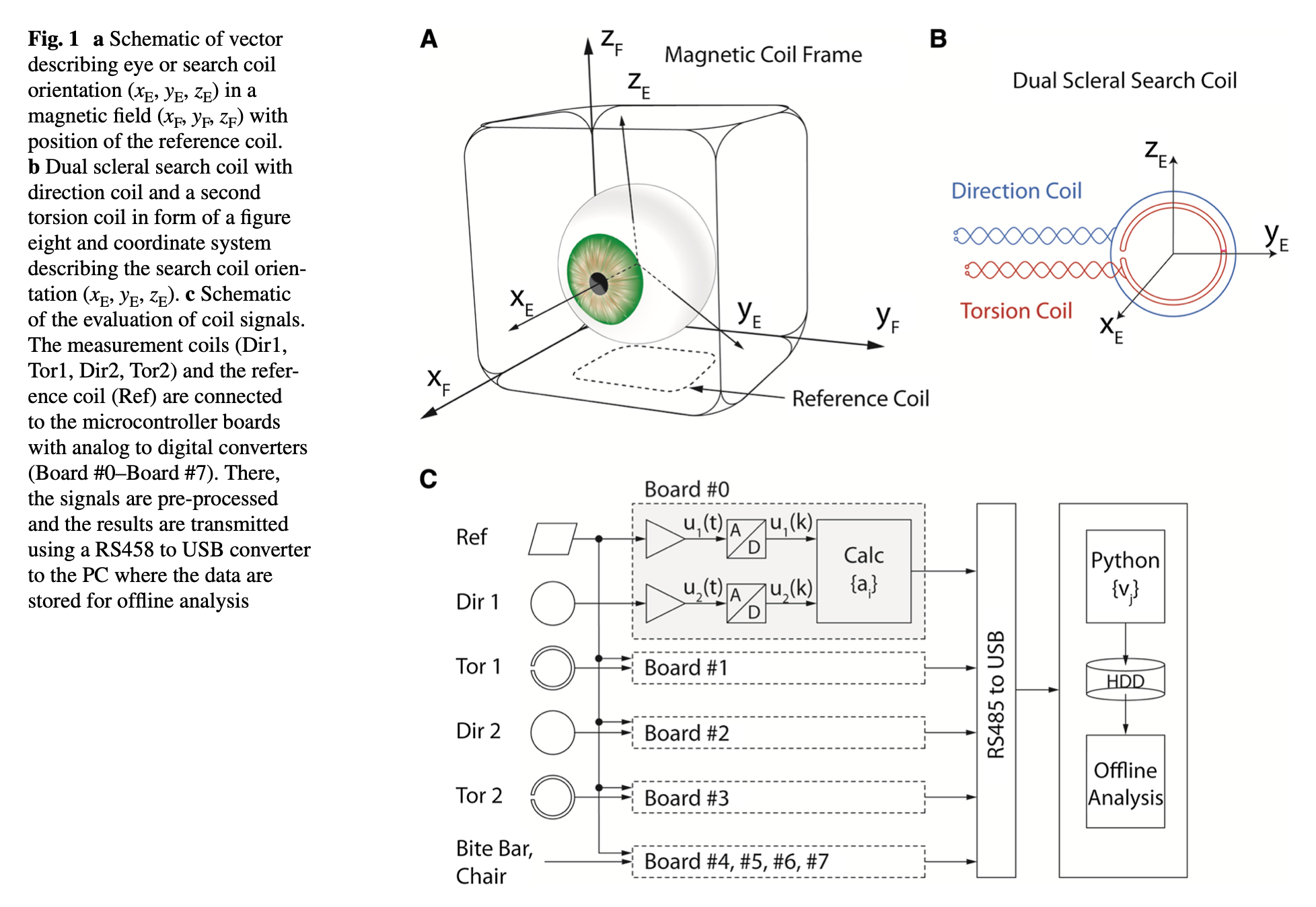
The Figure below, from Eibenberger and colleagues (Eibenberger et al. 2016), shows a schematic of this system, including the dual magnetic scleral search coil and the cubic magnetic field coil system.
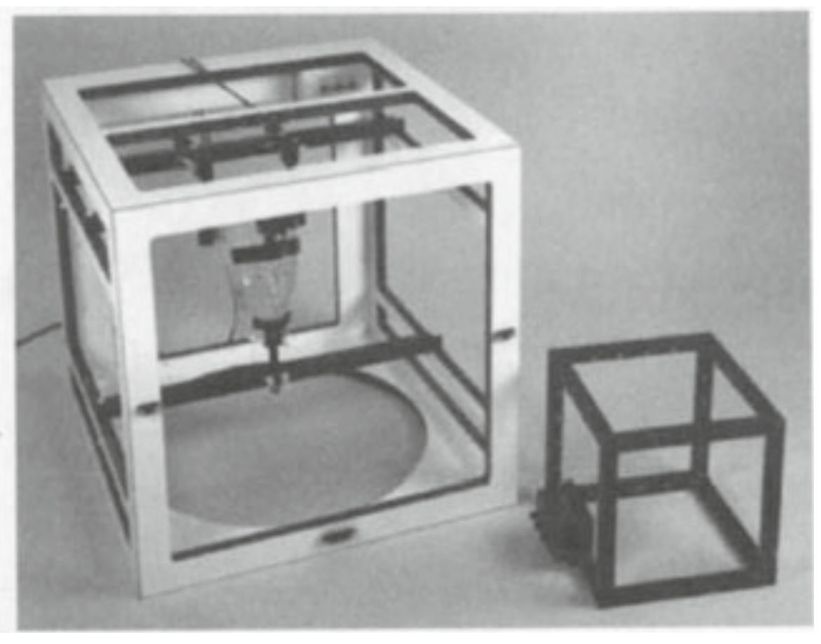
The Figure below, from Duchowski (Duchowski 2003), shows examples of cubic magnetic field coil frames for the head (left) and eyes (right).
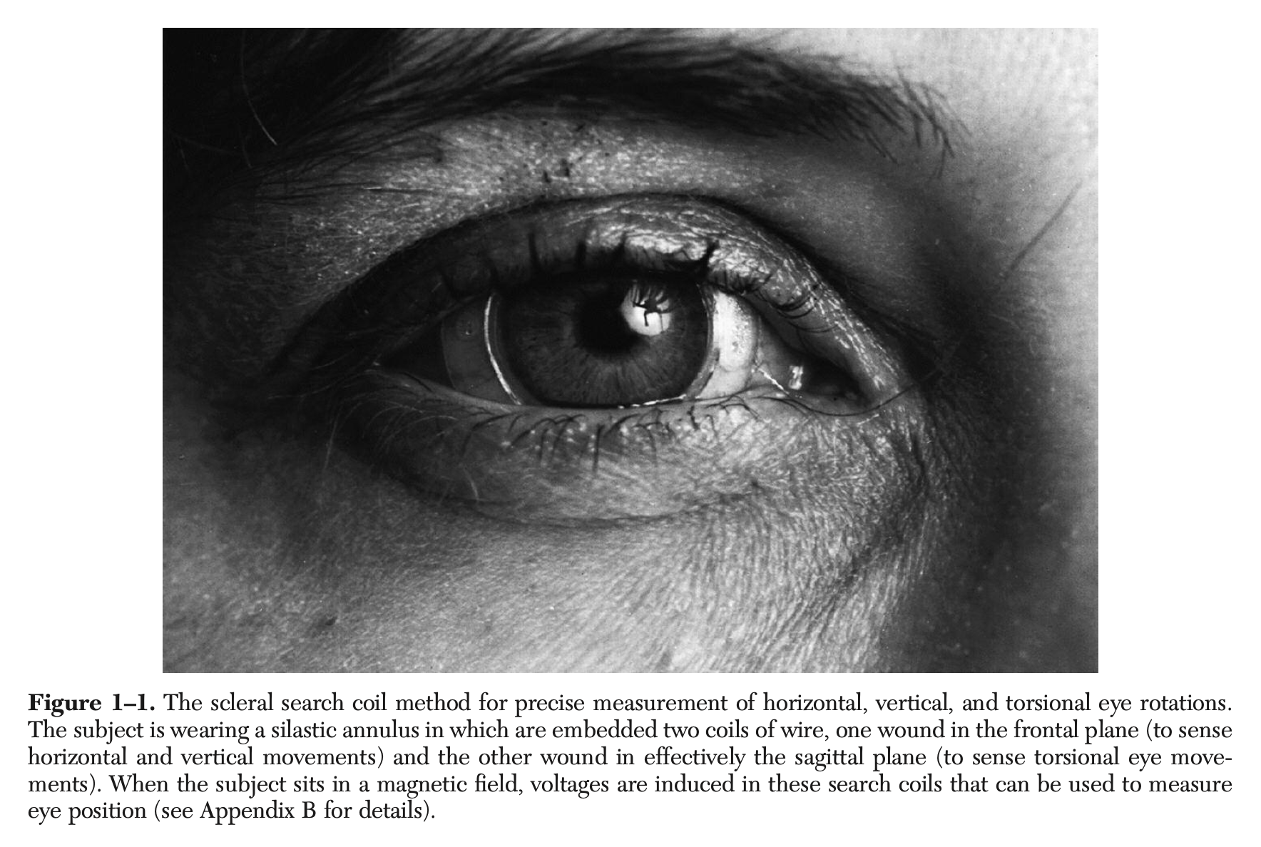
The Figure below, from Leigh and Zee (Leigh and Zee 2015), is a photograph of a patient’s eye on which a magnetic scleral search coil contact lens has been laced. Notice the very fine wire emerging from the contact lens at the patient’s medial canthus.
Subsequent modification to this system involved embedding a capacitor in the contact lens, enabling “wireless” use of the magnetic scleral search coil technique (Roberts et al. 2008). Scherer and colleagues (Scherer et al. 2011) explain this method as follows:
“The wireless scleral search coil is an annular contact lens that contains a small coil of fine wire and a capacitor embedded in a biocompatible polymer. Like the conventional ‘wired’ search coil, the lens is placed on the eye over the sclera near the corneal junction where it adheres by natural mild suction. While similar in many respects to a conventional wired scleral search coil system… the WSC [wireless scleral search coil] system uses a resonant scleral coil with no connecting wire. Instead, a transmitter sends a stream of pulses to the eye coil, and a receiver then detects the resonant oscillations re-radiated from the eye coil. The relative intensity of the signal, as received by sets of orthogonal receiver coils, determines the orientation of the eye coil. This approach retains the accuracy, precision, and high sampling rate of the traditional wired coil system, with added advantages of increased comfort to the subject and system portability” (Scherer et al. 2011).
This methodology is sometimes referred to as the “double magnetic induction” (DMI) technique (Bremen et al. 2007) since there is alternation between (1) delivery of a magnetic pulse from the frame in order to induce a current in the scleral search coil that charges the capacitor, and (2) discharge of the capacitor into the scleral search coil that creates a magnetic dipole which is detected by the electrical frame.
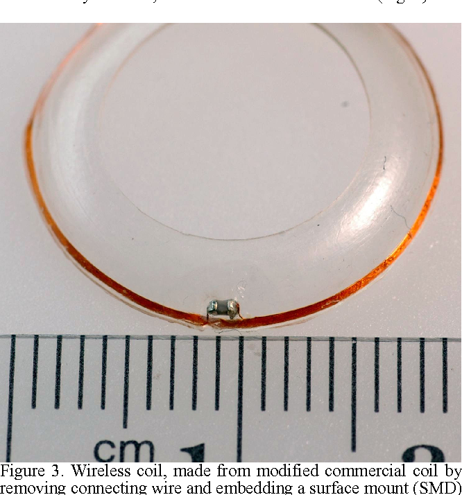
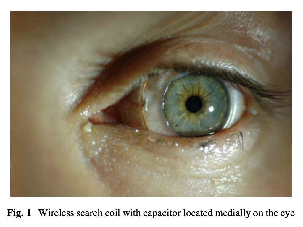
The Figure below, from Roberts and colleagues (Roberts et al. 2008), shows a contact lens with the embedded capacitor. The Figure below, from Scherer and colleagues (Scherer et al. 2011), shows this wireless contact lens worn by a patient.
 |
 |
The magnetic scleral search coil technique has the following characteristics, as described by Leigh and Zee:
“It allows measurement of eye rotations around all three axes, with a sensitivity of greater than 5 minutes of an arc (the standard deviation of system noise is typically less than 0.02 degree), a potential linear range of 360 degrees, a bandwidth of 0 Hz – 500 Hz, minimal drift, insensitivity to translation of the eye, and an unlimited field of view” (Leigh and Zee 2015).
These desirable features have not yet been surpassed by other techniques.
However, disadvantages of this technique include that it requires the patient to wear a contact lens, it is too cumbersome to be used for everyday clinical purposes, and its operation requires specialized training that most audiologists and ototechnicians never receive.
Equipment needed
Scleral search coil systems are usually constructed by engineers in research settings.
How to perform the test
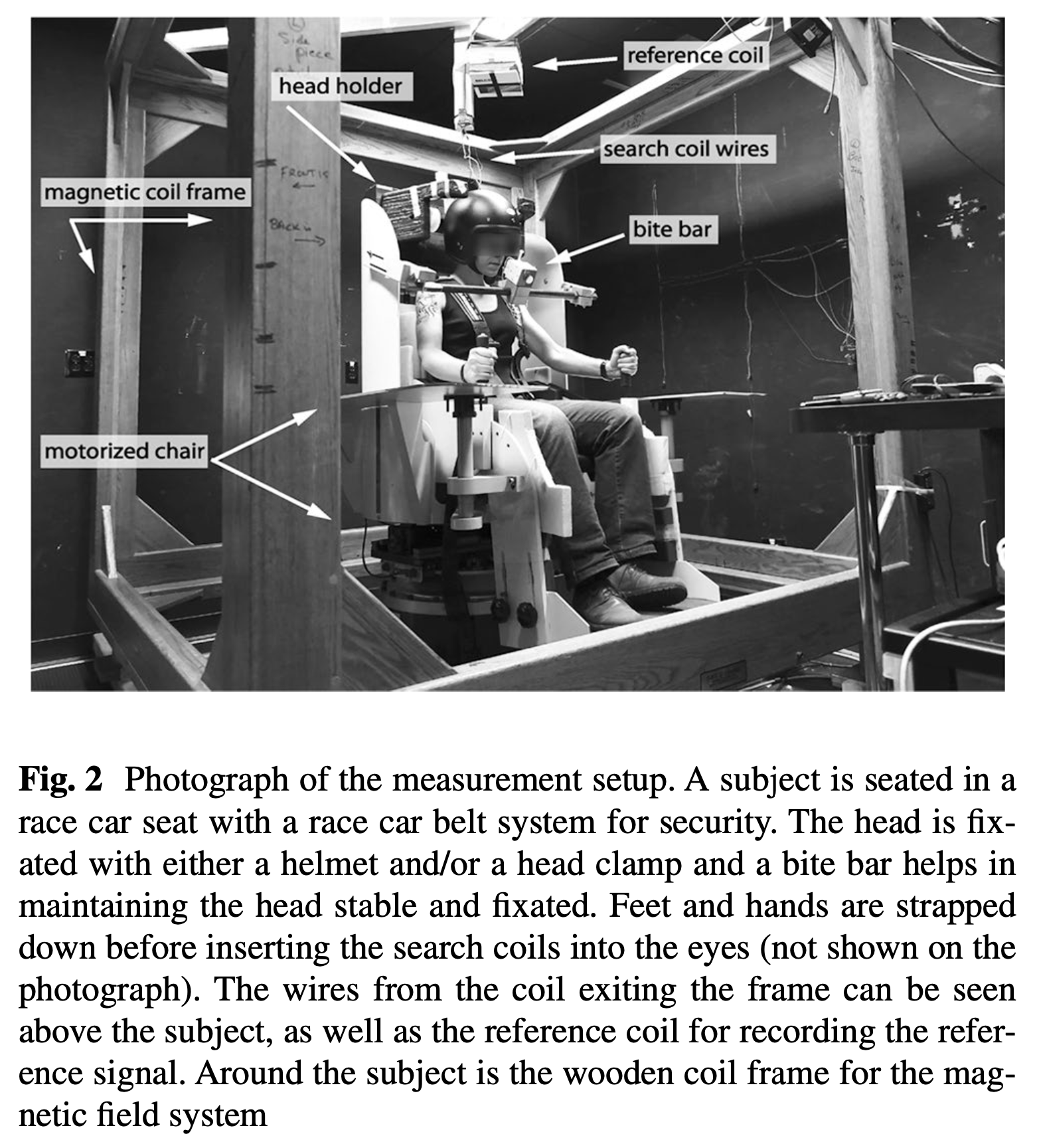
The cornea of the eye onto which the contact lens will be applied is anesthetized with oxyprocaine (Imai et al. 2005) or oxybuprocaine (Bremen et al. 2007). In many settings the position of the head is secured in order to isolate eye movements. For setups in which one needs to measure the position of an unsecured head, a search coil may also be applied to the patient’s head. The patient is then seated in such a way that the head is within the cubic magnetic field coil system.
The Figure below, from Eibenberger and colleagues (Eibenberger et al. 2016), shows the rather elaborate arrangement required for conducting a magnetic scleral search coil experiment.
Modifications
Given how cumbersome the arrangement is for a true dual magnetic scleral search coil system and double magnetic induction scleral search coil system, there have been attempts to develop simpler systems.
For example, Biancalana and Chessa (Biancalana and Chessa 2023) have developed a non-inducing magnetic contact lens — meaning that there is literally a static magnet embedded within the contact lens, as shown in the Figure below.
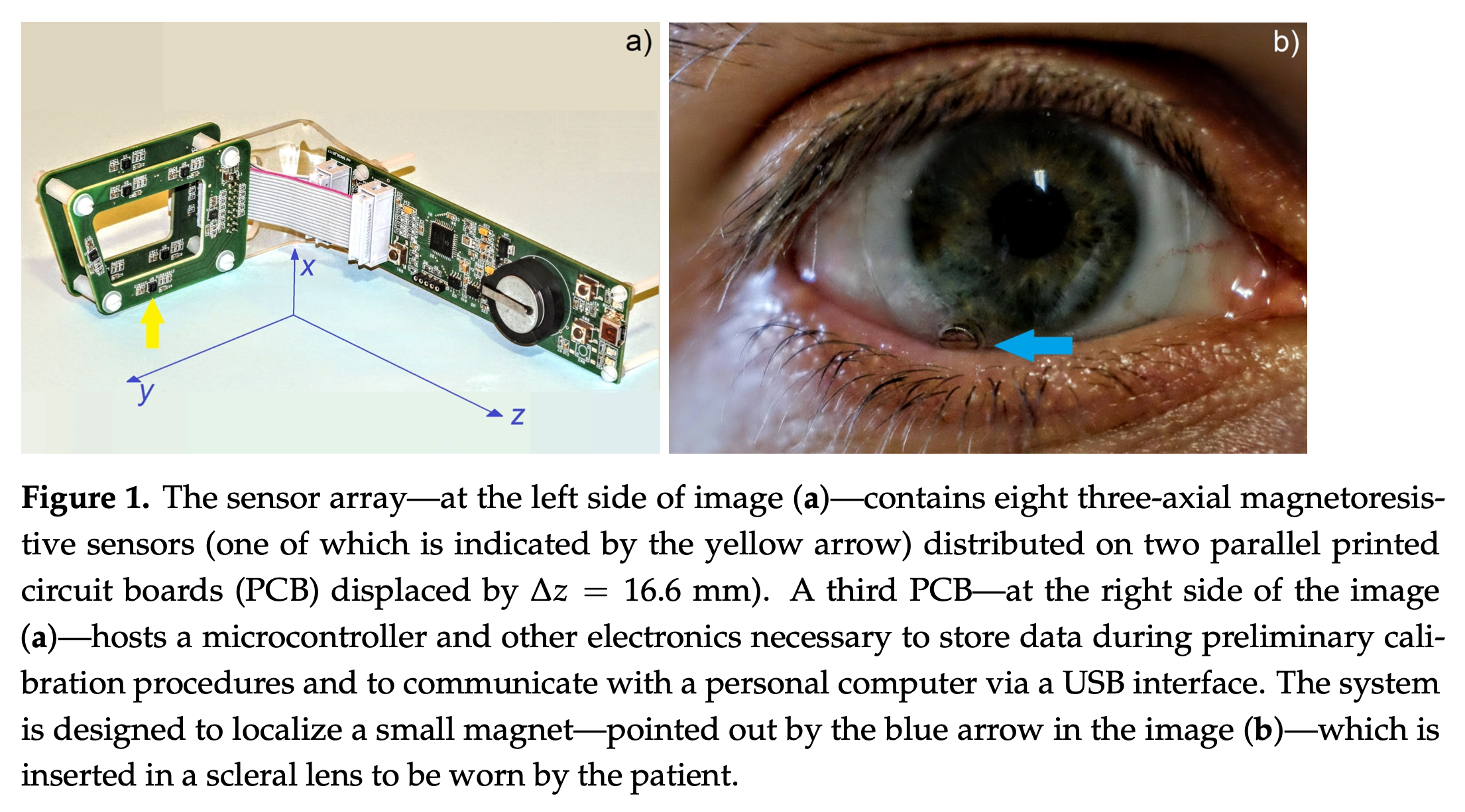
This method has not yet approached the accuracy of true scleral search coils.
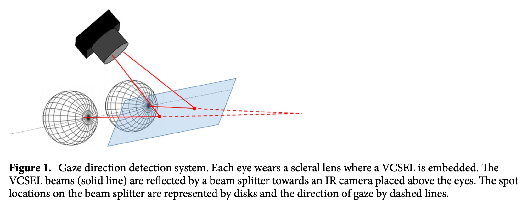
Another approach has been to maintain the foundation of the contact lens, but use tracking methodologies other than magnetism. For example, the Figure below, from Khaldi and colleagues (Khaldi et al. 2020), utilizes a vertical cavity surface-emitting laser embedded in a smart contact lens.
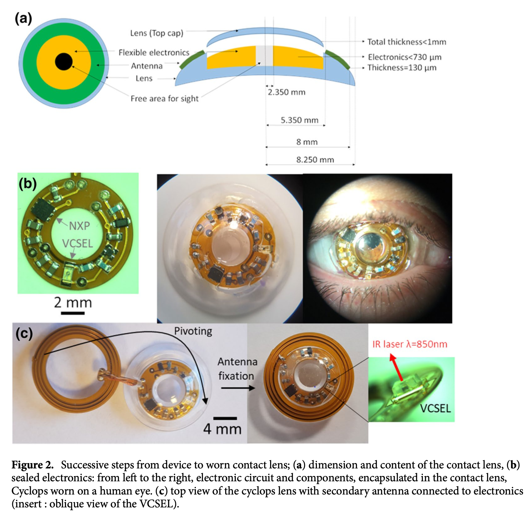
The emitted laser is reflected by a beam splitter and detected by a camera, as shown in the Figure below, from Khaldi and colleagues (Khaldi et al. 2020).
These alternative approaches hold promise, but have not yet surpassed the original magnetic scleral search coil technique.
References
Biancalana V, Chessa P (2023) A Non-Inductive Magnetic Eye-Tracker: From Dipole Tracking to Gaze Retrieval. Instruments, vol 7
Bremen P, Van der Willigen RF, Van Opstal AJ (2007) Applying double magnetic induction to measure two-dimensional head-unrestrained gaze shifts in human subjects. J Neurophysiol 98: 3759-69. doi: 10.1152/jn.00886.2007
Collewijn H, van der Mark F, Jansen TC (1975) Precise recording of human eye movements. Vision Res 15: 447-50. doi: 10.1016/0042-6989(75)90098-x
Collewijn H, Van der Steen J, Ferman L, Jansen TC (1985) Human ocular counterroll: assessment of static and dynamic properties from electromagnetic scleral coil recordings. Exp Brain Res 59: 185-96.
Duchowski AT (2003) Eye tracking methodology : theory and practice. Springer, New York
Eibenberger K, Eibenberger B, Roberts DC, Haslwanter T, Carey JP (2016) A novel and inexpensive digital system for eye movement recordings using magnetic scleral search coils. Med Biol Eng Comput 54: 421-30. doi: 10.1007/s11517-015-1326-3
Imai T, Sekine K, Hattori K, Takeda N, Koizuka I, Nakamae K, Miura K, Fujioka H, Kubo T (2005) Comparing the accuracy of video-oculography and the scleral search coil system in human eye movement analysis. Auris Nasus Larynx 32: 3-9. doi: 10.1016/j.anl.2004.11.009
Khaldi A, Daniel E, Massin L, Karnfelt C, Ferranti F, Lahuec C, Seguin F, Nourrit V, de Bougrenet de la Tocnaye JL (2020) A laser emitting contact lens for eye tracking. Sci Rep 10: 14804. doi: 10.1038/s41598-020-71233-1
Leigh RJ, Zee DS (2015) The neurology of eye movements, 5th edn. Oxford University Press, Oxford ; New York
Roberts D, Shelhamer M, Wong A A New “Wireless” Search-Coil System Eye Tracking Research and Applications Symposium, ETRA 2008, Savannah, Georgia 2008, pp 197-204
Robinson DA (1963) A Method of Measuring Eye Movement Using a Scleral Search Coil in a Magnetic Field. IEEE Trans Biomed Eng 10: 137-45.
Scherer MR, Shelhamer MJ, Schubert MC (2011) Characterizing high-velocity angular vestibulo-ocular reflex function in service members post-blast exposure. Exp Brain Res 208: 399-410. doi: 10.1007/s00221-010-2490-1
Shelhamer M, Roberts DC (2010) Chapter 6 – Magnetic scleral search coil. Handbook of Clinical Neurophysiology, vol 9. Elsevier, pp 80-87
![]()