By Marcello Cherchi, MD PhD
For patients
The pineal gland is a structure deep in the brain. If the pineal gland greatly enlarges (such as with a cyst or a tumor), then it can cause problems with eye movements, vision, walking and sometimes with hearing, and in such cases, a neurosurgeon may recommend removing the pineal mass. In cases where the pineal gland is affected by a non-malignant cyst that is not very large, then it is unlikely to be causing symptoms, and neurosurgeons will usually not recommend surgery.
For clinicians
Overview
Large pineal masses can have mechanical effects by narrowing or closing the cerebral aqueduct (resulting in obstructive hydrocephalus) and compressing the tectal plate (causing Parinaud syndrome and sometimes auditory symptoms). Non-malignant pineal cysts are frequently encountered as incidental findings on brain imaging. If a pineal mass (of any etiology) is causing mechanical compression, then neurosurgical intervention is indicated. In cases of non-malignant pineal cysts that are not causing mechanical compression, management is less clear; most clinicians will refrain from neurosurgical intervention.
Introduction
Large pineal masses can narrow or close the cerebral aqueduct (resulting in obstructive hydrocephalus) and compress the tectal plate (causing Parinaud syndrome and sometimes auditory symptoms), with corresponding abnormalities on physical examination, ocular motor examination, and audio-vestibular evaluation.
In routine clinical practice, the most commonly encountered pineal mass is a non-malignant pineal cyst found incidentally on brain imaging. Such pineal cysts usually remain stable in size; a small proportion of them (about 2.6% – 5.5%) enlarge. If a non-malignant pineal cyst is large enough to be causing obstructive hydrocephalus or Parinaud syndrome (dorsal midbrain syndrome), then neurosurgical intervention is indicated. If a non-malignant pineal cyst is not that large (and thus causing neither obstructive hydrocephalus nor Parinaud syndrome), then it is difficult to establish a causative relationship between the cyst and any symptoms, so management is less clear.
Many cases of pineal masses have been reported in the literature. Case series (whether prospective or retrospective) are more helpful in understanding the behavior of these lesions. Fortunately, there have been several informative case series of pineal masses (Fernandes et al. 2026; Gregory et al. 2011; Hajnsek et al. 2013; Hankinson et al. 2016; Hoehn et al. 2017; Májovský et al. 2017; Shields et al. 2017).
Epidemiology
Non-malignant pineal cysts are a common incidental radiologic finding.
Fernandes and colleagues (Fernandes et al. 2026) note that literature cites prevalence rates of non-malignant pineal cysts in the general population as ranging widely from 1.4% to 37.5%. Pineal cysts are more common in women. Of all pineal cysts, about 5% increase in size; the remainder are static.
Májovský and colleagues (Májovský et al. 2017) prospective studied 110 patients with pineal cysts, with a mean follow-up time of 79.2 months. They reported that the pineal cyst increased in size in 6 patients (5.5%), decreased in size in 9 patients (8.2%), and were stable (unchanged in size) in the remaining 95 (86.3%).
Al-Holou and colleagues (Al-Holou et al. 2011) studied 48,417 consecutive adults (over 18 years) who underwent brain MRI over a 12-year period. They found pineal cysts measuring 5 mm or larger in greatest dimensions in 478 patients (1.0%), of which 162 (33.9%) were male and 316 (66.1%) were female. Of 151 patients with subsequent brain MRIs (done at a mean interval of 3.4 years from the index study), 4 (2.6%) increased in size, 23 (15.2%) decreased in size, and 124 (82.1%) pineal cysts remained stable.
Al-Holou and colleagues (Al-Holou et al. 2011) then combined MRI data from the same 48,417 consecutive adults (over 18 years) with 14,516 consecutive children (≤8 years) on whom they had previously reported (Al-Holou et al. 2009), and arrived at the age-based histogram of prevalence of pineal cysts shown in the Figure below.
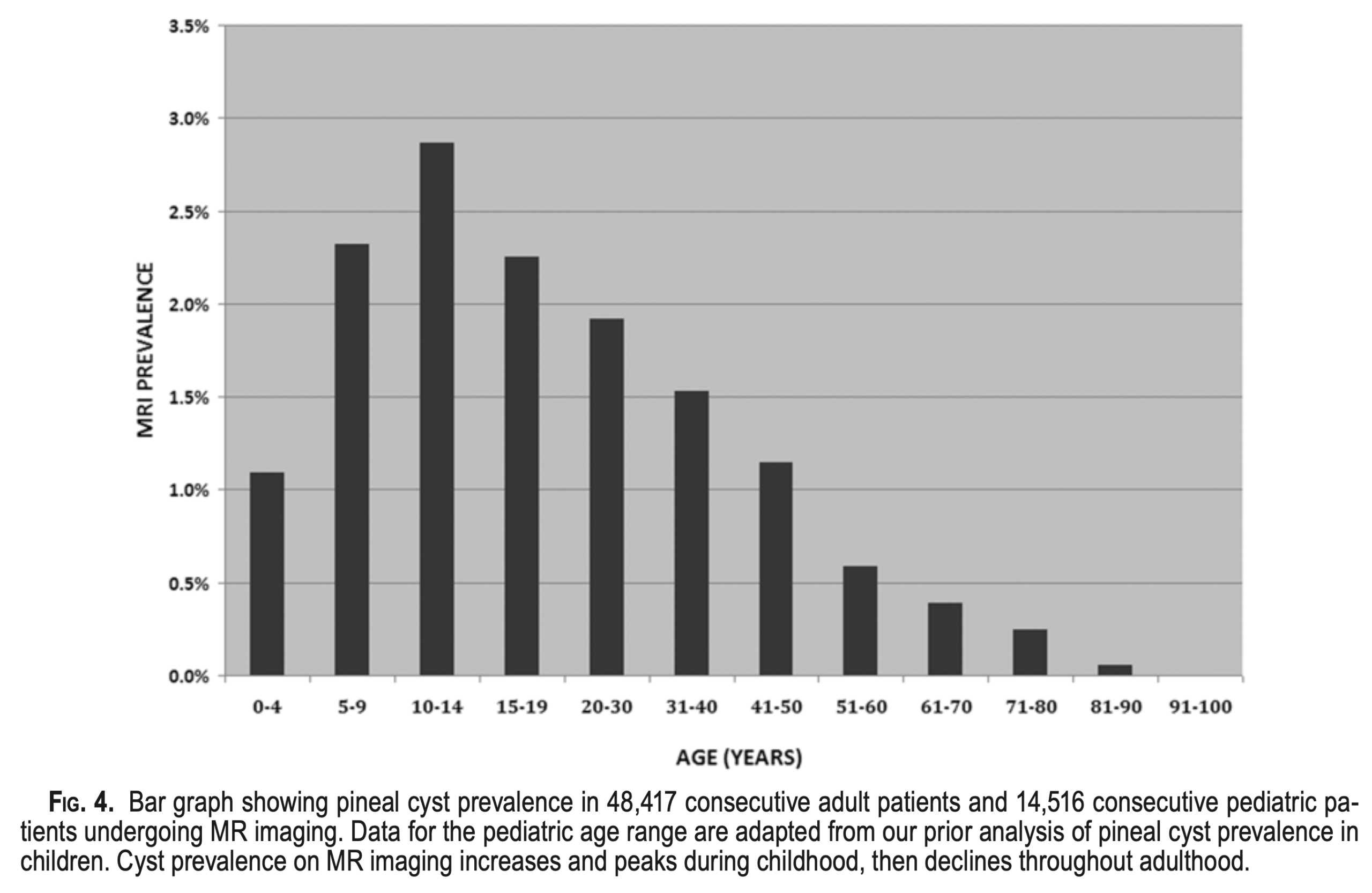
Figure : Histogram showing the prevalence of pineal cysts in 48,417 consecutive adult patients and 14,516 consecutive pediatric patients undergoing MRI. From Al-Holou et al (Al-Holou et al. 2011).
Genetics
Until recently there were no known genetic factors predisposing to the development of non-malignant pineal cysts. With the advent of whole exome sequencing, some research suggests that several hundred genetic variants may be associated with non-malignant pineal cysts (Yan et al. 2021). While this is interesting, there is not yet any practical consequence, so genetic testing currently plays no role in the routine evaluation of non-malignant pineal cysts.
Some malignant pineal masses do have known genetic associations. For example, a malignant pineal mass occurring in a patient with bilateral retinoblastomas (OMIM 180200) is associated with a mutation on chromosome 13q14.2 encoding retinoblastoma transcriptional corepressor 1 (RB1) (OMIM 614041).
Pathophysiological mechanism of disease
Pineal masses (of any etiology) can provoke symptoms when they are large enough to cause the mechanical effects of (1) blocking the quadrigeminal cistern, thereby limiting the flow through the cerebral aqueduct and leading to obstructive hydrocephalus, and/or (2) compressing the tectal plate.
The various etiologies of pineal masses vary in prevalence, and case series report different rates.
For example, Hajnsek and colleagues (Hajnsek et al. 2013) studied a retrospective series of 84 patients who underwent surgery for pineal masses. They reported the following histopathologies:
- Pineal gland cysts in 80%
- Pineocytomas in 16%
- Pineoblastomas in 3%
- Teratoma in 1%
In contrast, Shields and colleagues (Shields et al. 2017) retrospectively reviewed 40 cases of Parinaud syndrome. Twelve of these cases were due to pineal tumors, and the histopathologies were:
- Germinoma in 4 (25%)
- Pineoblastoma in 2 (17%)
- Meningioma in 2 (17%)
- Astrocytoma in 1 (8%)
- Cavernoma in 1 (8%)
- Choriocarcinoma in 1 (8%)
- Diffuse B-cell lymphoma in 1 (8%)
Clinical presentation
A non-malignant pineal cyst is a common incidental finding on brain imaging (see the earlier section on epidemiology). Incidentally discovered pineal cysts are rarely the cause of symptoms (Al-Holou et al. 2011; Choy et al. 2011; Di Costanzo et al. 1993; Pu et al. 2007).
When a pineal mass (of any etiology) causes obstructive hydrocephalus, common presenting symptoms are those of hydrocephalus in general, including nausea, vomiting and visual abnormalities (Upadhyayula et al. 2023), including from ocular motor cranial nerve palsies.
Májovský and colleagues (Májovský et al. 2017) prospectively studied 110 patients with pineal cysts who were referred for neurosurgical consultation, and reported the following presenting symptoms:
- Tension headache in 62.7%
- Vertigo in 16.4%
- Migraine in 12.7%
- Syncope in 10.9%
- Nausea in 8.2%
- Diplopia 8.2%
Májovský and colleagues (Májovský et al. 2017) followed these 110 patients over a mean follow-up time of 79.2 months and reported that symptoms worsened in 17 (15.5%), improved in 13 (11.8%) and remained stable in 81 (73.6%).
If a pineal mass compresses the inferior colliculi, then various auditory symptoms may result, including:
- Hearing loss (Gaspar et al. 2003; Islam et al. 2002; Mizobuchi et al. 2021; Woo et al. 2013)
- Word deafness (Joswig et al. 2015; Masuda et al. 2000)
- Tinnitus (Missori et al. 1995)
Physical examination
If a pineal mass is causing hydrocephalus, the ophthalmoscopic examination may show papilledema. Hankinson and colleagues (Hankinson et al. 2016) studied a case series of 29 children and reported that 69% exhibited papilledema.
Ocular motor examination
Pineal masses often manifest with Parinaud syndrome (also known as dorsal midbrain syndrome), whose classic presentation comprises the triad of conjugate upgaze paralysis, convergence-retraction nystagmus and light-near dissociation. However, Shields and colleagues (Shields et al. 2017) report that Parinaud syndrome presents with the full triad of signs in only 65% of cases. Hankinson and colleagues (Hankinson et al. 2016) studied a case series of 29 children and reported that 75% exhibited “partial or complete Parinaud’s syndrome.”
The upgaze paresis and convergence-retraction nystagmus can be elicited by vertical optokinetic stimulation (horizontal stripes moving vertically). This is one of the few instances in which it is helpful to use an optokinetic drum oriented such that the axis of rotation is parallel to the earth’s surface during ocular motor examination.
Less common manifestations in Parinaud syndrome include upper eyelid retraction (Collier’s sign), diplopia, visual field defects and ataxia (Orvis 2024). Gregory and colleagues report impaired downgaze and convergence insufficiency (Gregory et al. 2011). Hoehn and colleagues report exotropia (Hoehn et al. 2017).
Testing: auditory
If a pineal mass compresses the inferior colliculi, then various auditory symptoms may result, such as hearing loss, word deafness and tinnitus.
Gaspar and colleagues (Gaspar et al. 2003) report the case of a 12-year-old boy with a pineal germ cell tumor that was compressing the inferior colliculi. Audiometry showed approximately symmetrical sensorineural hearing loss (greater in the high frequencies than the middle frequencies) and complete word deafness, as shown in the Figure below.
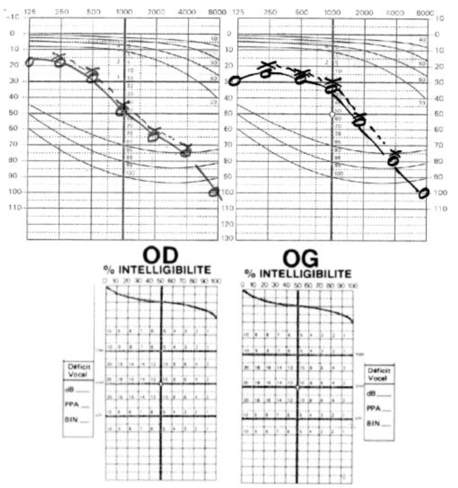
Figure : Case of a 12-year-old boy with pineal germ cell tumor compressing the inferior colliculi. Audiometry shows approximately symmetrical high greater than middle frequency sensorineural hearing loss, and complete word deafness. From Gaspar et al (Gaspar et al. 2003).
Compression of the inferior colliculi by a pineal mass can also cause abnormalities in auditory brainstem evoked responses (ABR). For example, Mizobuchi and colleagues (Mizobuchi et al. 2021) report a case of a patient pineal glial cyst compressing the inferior colliculi, whose auditory brainstem evoked responses showed an absence of wave V, as displayed in the Figure below.
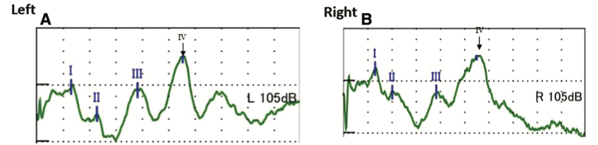
Figure : Auditory brainstem evoked response in a patient with a pineal glial cyst compressing the inferior colliculi. Note the absence of wave V bilaterally. From Mizobuchi et al (Mizobuchi et al. 2021).
Woo and colleagues (Woo et al. 2013) reported the case of a patient with a pineal ganglioglioma compressing the inferior colliculi. In this case wave V was preserved, but the wave III-V interpeak interval was prolonged.
Testing: vestibular
Baloh and colleagues (Baloh et al. 1985) reported a series of patients with Parinaud’s syndrome, of which 5 were cases of pineal tumors.
The Figure below, from Baloh and colleagues (Baloh et al. 1985), shows impairment of vertical saccades in a 15-year-old boy with a pineal dysgerminoma.
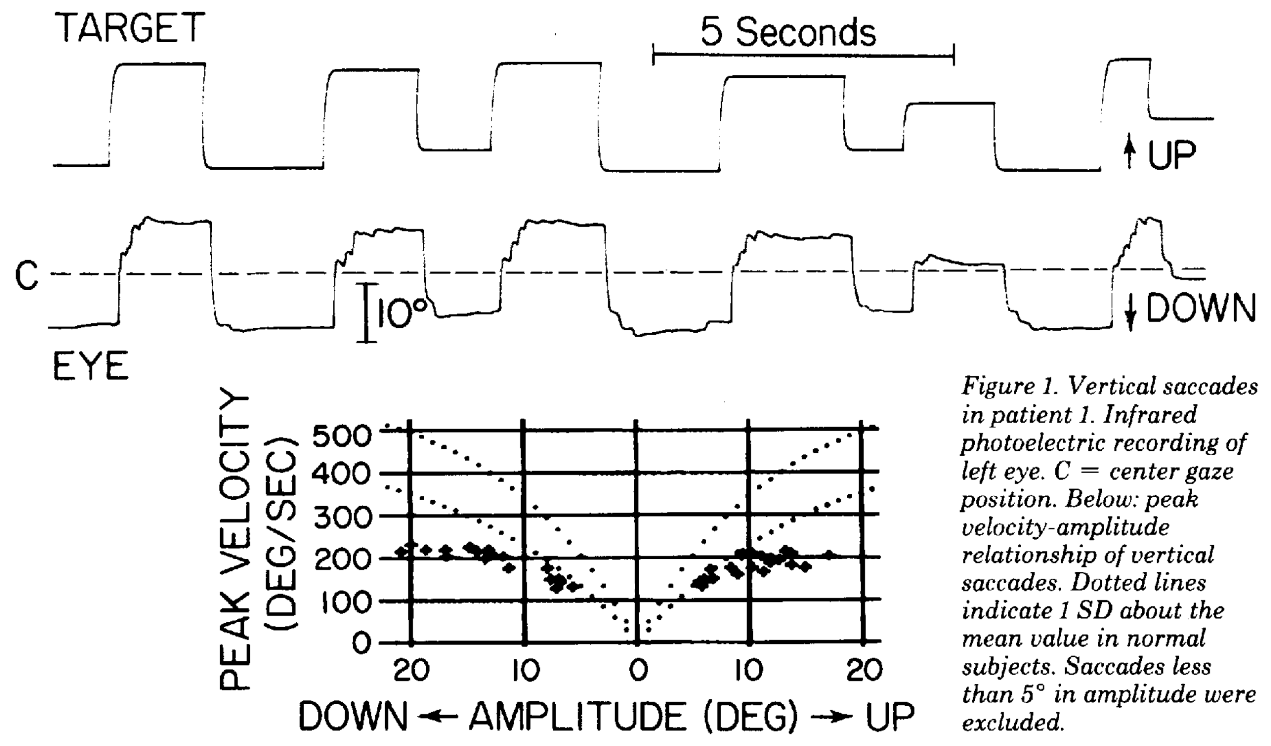
Figure : Ocular motor tracings of a 15-year-old boy with pineal dysgerminoma. Note that upward saccades are hypometric by more than 1 standard deviation. From Baloh et al (Baloh et al. 1985).
Eliciting convergence-retraction nystagmus can be done very effectively with a projected optokinetic stimulus, as shown in the Figure below from Garbutt and Harris (Garbutt and Harris 2000).
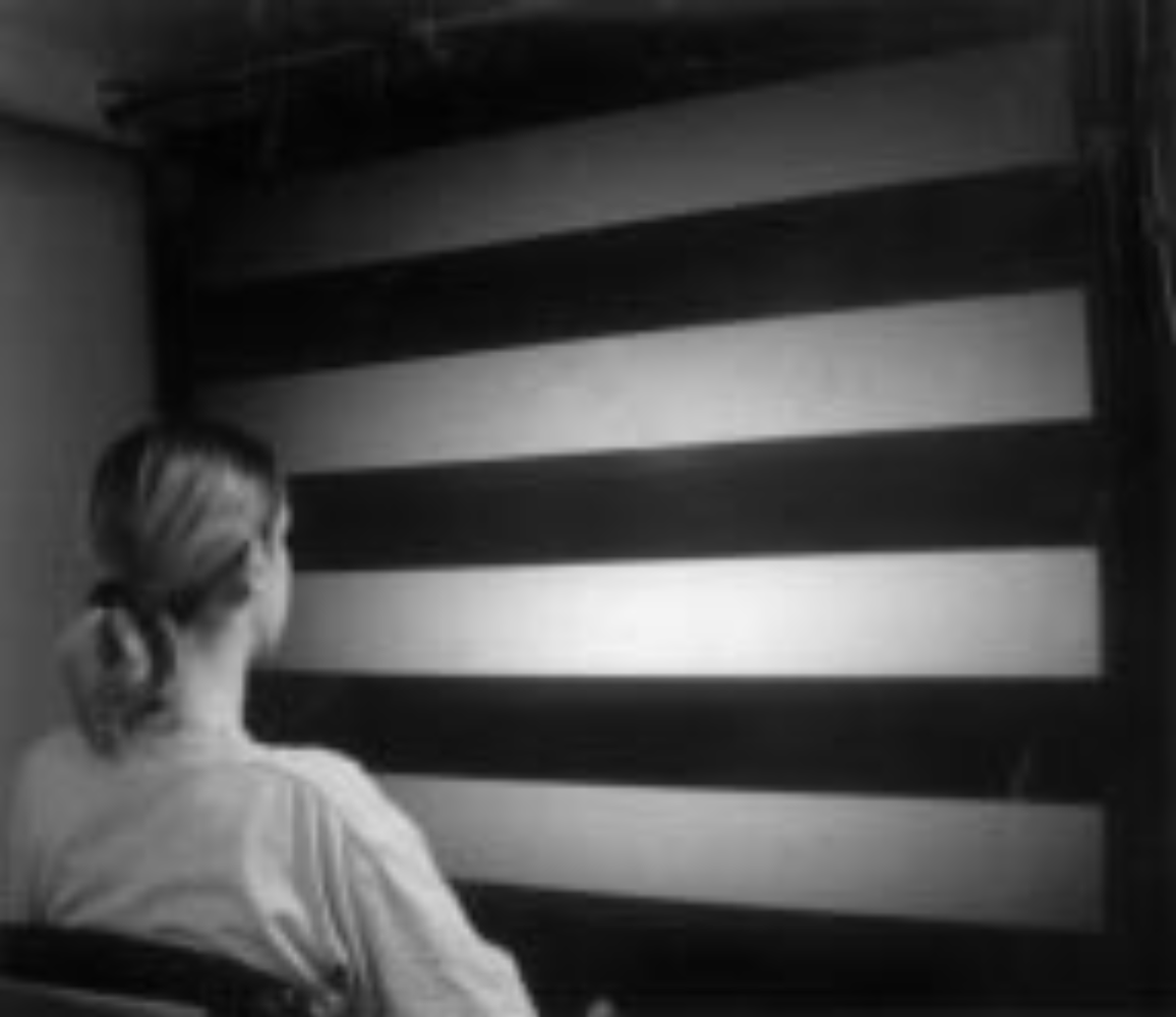
Figure : Projection of a vertically moving optokinetic stimulus. From Garbutt and Harris (Garbutt and Harris 2000).
Imaging
The Figure below, from Aaroe and colleagues (Aaroe et al. 2021), shows an MRI from a patient whose pineal mass is a primary pineal melanoma.
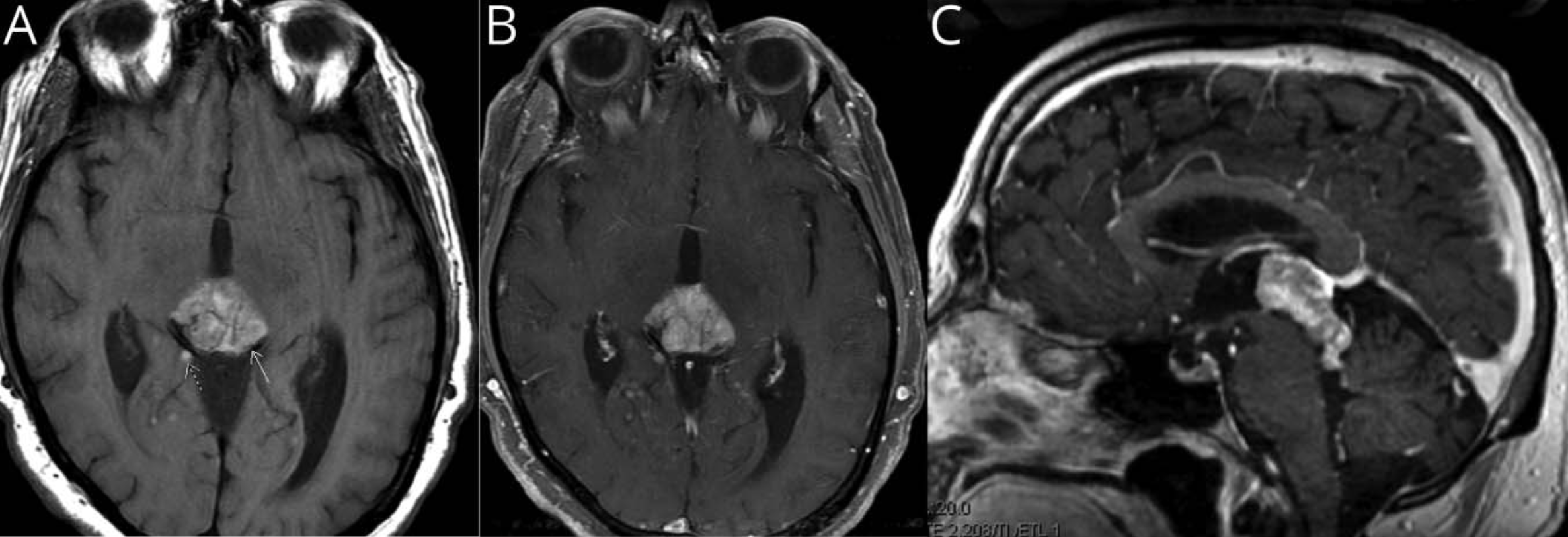
Figure : MRI of a patient with a primary pineal melanoma. Panel (A) shows a hyperintense pineal region mass. Panel (B) shows enhancement of this mass, as well as enhancement of numerous foci in the subarachnoid space. Panel (C) shows high signal intensity on T1-weighted images. From Aaroe et al (Aaroe et al. 2021).
Histopathology
Pineal masses can result from a range of etiologies, including:
- Cyst (Bosnjak et al. 2009; Fleck et al. 2022; Gotz Wieckowska et al. 2017; Lukewich et al. 2020; Woodward et al. 2020)
- Arachnoid cyst (Gonzalez et al. 2007)
- Epidermoid cyst (Konovalov et al. 1999)
- Ganglioglioma (Woo et al. 2013)
- Germinoma (Gaspar et al. 2003; Gurusamy et al. 2022; Islam et al. 2002; Joswig et al. 2015; Nishibayashi et al. 2005; Prasetio et al. 2022; Wong et al. 2023; Yang et al. 2004)
- Glial cyst (Mizobuchi et al. 2021)
- Granular cell astrocytoma (El Asri et al. 2015)
- Hemangioblastoma (Isolan et al. 2007)
- Melanoma (Aaroe et al. 2021)
- Metastatic small cell carcinoma (Kashiwagi et al. 1989)
- Neuroendocrine carcinoma (Hakar et al. 2017)
- Papillary tumor (Boco et al. 2008; Mobark et al. 2022)
- Paraganglioma (Reithmeier et al. 2000)
- Pineoblastoma (Saito et al. 2002; Yiğit and Gökoğlu 2025)
- Pineocytoma (Allmer and Golis 2001)
- Rosette-forming glioneuromal tumor (Solis et al. 2011)
- Sarcoidosis (Adel et al. 2021)
- Teratoma (Lewis et al. 1963)
- Trilateral retinoblastoma (Bejjani et al. 1996; Cho et al. 2002)
- Indeterminate or undifferentiated tissue (Shrateh et al. 2023)
Differential diagnosis
The main differential diagnosis of symptomatic pineal cysts includes other causes of hydrocephalus, and other causes of Parinaud syndrome (such as intrinsic lesions of the tectal plate).
Management
When a pineal mass — including a non-malignant pineal cyst — is large enough to cause obstructive hydrocephalus or Parinaud syndrome, then neurosurgical resection is indicated.
For patients who have pineal cysts that are not causing obstructive hydrocephalus or Parinaud syndrome, yet still have symptoms, it is difficult to establish a causative relationship between the pineal mass and the symptoms with confidence, so management is unclear.
Kalani and colleagues (Kalani et al. 2015) retrospectively described a series of 18 patients (14 female, 4 male; mean age 24 years, range 4 – 47 years) who underwent neurosurgical resection of benign pineal cysts (preoperative cyst diameter 1.5 cm, with a range of 0.9 – 2.2 cm) that were not causing obstructive hydrocephalus or Parinaud syndrome. The presenting symptoms in these 18 patients were:
- Headache in 17 (94%)
- Visual disturbances in 10 (56%)
- Gait instability in 5 (28%)
- Dizziness in 5 (28%)
- Recurrent syncope in 2 (11%)
- Hypersomnolence in 1 (6%)
Kalani and colleagues (Kalani et al. 2015) followed these patients for a mean of 19.1 months (ranging from postoperative to 71 months) and reported that 17 (94%) patients had “resolution of improvement in their presenting symptoms.”
Prognosis
Hoehn and colleagues (Hoehn et al. 2017) described a series of pediatric patients with Parinaud syndrome from pineal tumors. Out of 17 patients who underwent surgery, 7 (41%) exhibited postoperative improvement in the ocular motor abnormalities, but only 2 (12%) exhibited complete resolution.
When a pineal mass causes auditory symptoms due to compression of the inferior colliculi, treatment of the mass may bring about improvement in auditory symptoms (Gaspar et al. 2003; Joswig et al. 2015; Mizobuchi et al. 2021; Woo et al. 2013), as well as in audiometric results such as pure tone audiometry (Gaspar et al. 2003; Mizobuchi et al. 2021; Woo et al. 2013), word recognition (Gaspar et al. 2003; Woo et al. 2013), and auditory brainstem evoked responses (Islam et al. 2002).
References
Aaroe AE, Glitza Oliva IC, Al-Zubidi N, Nader ME, Kaya D, Ferguson SD, Wang C, Fuller G, Harrison RA (2021) Pearls & Oy-sters: Primary Pineal Melanoma With Leptomeningeal Carcinomatosis. Neurology 97: 248-250. doi: 10.1212/wnl.0000000000012121
Adel K, Bennabi W, Aichaoui F, Meziani S, Morsli A (2021) Sarcoidosis of the pineal region, dorsal mesencephalon, and periaqueductal region: Case report and systematic review. Qatar Med J 2021: 29. doi: 10.5339/qmj.2021.29
Al-Holou WN, Garton HJ, Muraszko KM, Ibrahim M, Maher CO (2009) Prevalence of pineal cysts in children and young adults. Clinical article. J Neurosurg Pediatr 4: 230-6. doi: 10.3171/2009.4.PEDS0951
Al-Holou WN, Terman SW, Kilburg C, Garton HJ, Muraszko KM, Chandler WF, Ibrahim M, Maher CO (2011) Prevalence and natural history of pineal cysts in adults. J Neurosurg 115: 1106-14. doi: 10.3171/2011.6.JNS11506
Allmer DM, Golis TA (2001) Dorsal midbrain syndrome secondary to a pineocytoma. Optometry 72: 234-8.
Baloh RW, Furman JM, Yee RD (1985) Dorsal midbrain syndrome: clinical and oculographic findings. Neurology 35: 54-60. doi: 10.1212/wnl.35.1.54
Bejjani GK, Donahue DJ, Selby D, Cogen PH, Packer R (1996) Association of a suprasellar mass and intraocular retinoblastoma: a variant of pineal trilateral retinoblastoma? Pediatr Neurosurg 25: 269-75. doi: 10.1159/000121138
Boco T, Aalaei S, Musacchio M, Byrne R, Cochran E (2008) Papillary tumor of the pineal region. Neuropathology 28: 87-92. doi: 10.1111/j.1440-1789.2007.00832.x
Bosnjak J, Budisić M, Azman D, Strineka M, Crnjaković M, Demarin V (2009) Pineal gland cysts–an overview. Acta Clin Croat 48: 355-8.
Cho EY, Suh YL, Shin HJ (2002) Trilateral retinoblastoma: a case report. J Korean Med Sci 17: 137-40. doi: 10.3346/jkms.2002.17.1.137
Choy W, Kim W, Spasic M, Voth B, Yew A, Yang I (2011) Pineal cyst: a review of clinical and radiological features. Neurosurg Clin N Am 22: 341-51, vii. doi: 10.1016/j.nec.2011.06.001
Di Costanzo A, Tedeschi G, Di Salle F, Golia F, Morrone R, Bonavita V (1993) Pineal cysts: an incidental MRI finding? J Neurol Neurosurg Psychiatry 56: 207-8. doi: 10.1136/jnnp.56.2.207
El Asri AC, Baallal H, Zoubeir Y, Sinaa M, Albouzidi A, Gazzaz M, Akhaddar A, Boucetta M, El Mostarchid B (2015) Diagnosis and management challenge of a granular cell astrocytoma of the pineal region: case report. J Neurosurg Pediatr 15: 506-9. doi: 10.3171/2014.10.Peds13388
Fernandes FW, Saryyeva A, Hermann EJ, Nakamura M, Krauss JK (2026) Pineal cyst surgery beyond morphology: a critical evaluation of a consecutive surgical series. Acta Neurochir (Wien) 168. doi: 10.1007/s00701-026-06845-z
Fleck S, Damaty AE, Lange I, Matthes M, Rafaee EE, Marx S, Baldauf J, Schroeder HWS (2022) Pineal cysts without hydrocephalus: microsurgical resection via an infratentorial-supracerebellar approach-surgical strategies, complications, and their avoidance. Neurosurg Rev 45: 3327-3337. doi: 10.1007/s10143-022-01831-2
Garbutt S, Harris CM (2000) Abnormal vertical optokinetic nystagmus in infants and children. Br J Ophthalmol 84: 451-5. doi: 10.1136/bjo.84.5.451
Gaspar N, Verschuur A, Mercier G, Couanet D, Sainte-Rose C, Brugières L (2003) Reversible hearing loss associated with a malignant pineal germ cell tumor. Case report. J Neurosurg 99: 587-90. doi: 10.3171/jns.2003.99.3.0587
Gonzalez C, Seth RK, Ramos-Esteban JC (2007) Change in head posture and character of nystagmus in a patient with neurological upbeat nystagmus. Binocul Vis Strabismus Q 22: 179-84.
Gotz Wieckowska A, Glowka L, Brazert A, Pawlak M (2017) Ophthalmological symptoms in children with intracranial cysts. Sci Rep 7: 13630. doi: 10.1038/s41598-017-13266-7
Gregory ME, Rahman MQ, Cleary M, Weir CR (2011) Dorsal midbrain syndrome with loss of motor fusion: a rare association. Strabismus 19: 17-20. doi: 10.3109/09273972.2010.545468
Gurusamy SM, Yaakub A, Wan Hitam WH, Ahmad Maulana SA, Wan Ibrahim WZ (2022) Pineal Gland Tumour With Drop Metastases: A Case Report. Cureus 14: e29855. doi: 10.7759/cureus.29855
Hajnsek S, Paladino J, Gadze ZP, Nanković S, Mrak G, Lupret V (2013) Clinical and neurophysiological changes in patients with pineal region expansions. Coll Antropol 37: 35-40.
Hakar M, Chandler JP, Bigio EH, Mao Q (2017) Neuroendocrine carcinoma of the pineal parenchyma. The first reported case. J Clin Neurosci 35: 68-70. doi: 10.1016/j.jocn.2016.09.024
Hankinson EV, Lyons CJ, Hukin J, Cochrane DD (2016) Ophthalmological outcomes of patients treated for pineal region tumors. J Neurosurg Pediatr 17: 558-63. doi: 10.3171/2015.10.Peds15415
Hoehn ME, Calderwood J, O’Donnell T, Armstrong GT, Gajjar A (2017) Children with dorsal midbrain syndrome as a result of pineal tumors. J aapos 21: 34-38. doi: 10.1016/j.jaapos.2016.09.024
Islam MS, Asano K, Tabata H, Ohkuma H, Suzuki S (2002) Pineal region tumor manifesting initially as hearing impairment. Neurol Med Chir (Tokyo) 42: 301-4. doi: 10.2176/nmc.42.301
Isolan GR, Krayenbühl N, Mahmoud M, Al-Mefty O (2007) A hemangioblastoma in the pineal region: case report. Neurosurgery 61: E423; discussion E423. doi: 10.1227/01.Neu.0000255516.12085.Bb
Joswig H, Schönenberger U, Brügge D, Richter H, Surbeck W (2015) Reversible pure word deafness due to inferior colliculi compression by a pineal germinoma in a young adult. Clin Neurol Neurosurg 139: 62-5. doi: 10.1016/j.clineuro.2015.08.034
Kalani MY, Wilson DA, Koechlin NO, Abuhusain HJ, Dlouhy BJ, Gunawardena MP, Nozue-Okada K, Teo C (2015) Pineal cyst resection in the absence of ventriculomegaly or Parinaud’s syndrome: clinical outcomes and implications for patient selection. J Neurosurg 123: 352-6. doi: 10.3171/2014.9.JNS141081
Kashiwagi S, Hatano M, Yokoyama T (1989) Metastatic small cell carcinoma to the pineal body: case report. Neurosurgery 25: 810-3. doi: 10.1097/00006123-198911000-00020
Konovalov AN, Spallone A, Pitzkhelauri DI (1999) Pineal epidermoid cysts: diagnosis and management. J Neurosurg 91: 370-4. doi: 10.3171/jns.1999.91.3.0370
Lewis I, Baxter DW, Stratford JG (1963) Atypical Teratomas of the Pineal. Can Med Assoc J 89: 103-10.
Lukewich MK, Alshafai L, Micieli JA (2020) Spontaneous Resolution of Dorsal Midbrain Syndrome Caused by a Pineal Cyst. World Neurosurg 136: 289-292. doi: 10.1016/j.wneu.2020.01.144
Májovský M, Netuka D, Beneš V (2017) Conservative and Surgical Treatment of Patients with Pineal Cysts: Prospective Case Series of 110 Patients. World Neurosurg 105: 199-205. doi: 10.1016/j.wneu.2017.05.155
Masuda S, Takeuchi K, Tsuruoka H, Ukai K, Sakakura Y (2000) Word deafness after resection of a pineal body tumor in the presence of normal wave latencies of the auditory brain stem response. Ann Otol Rhinol Laryngol 109: 1107-12. doi: 10.1177/000348940010901204
Missori P, Delfini R, Cantore G (1995) Tinnitus and hearing loss in pineal region tumours. Acta Neurochir (Wien) 135: 154-8. doi: 10.1007/bf02187761
Mizobuchi Y, Shimada A, Nakajima K, Kagusa H, Takagi Y (2021) Reversible Hearing Impairment Due to Inferior Colliculi Compression by a Pineal Glial Cyst. NMC Case Rep J 8: 79-84. doi: 10.2176/nmccrj.cr.2020-0123
Mobark NA, Alharbi M, Alotabi F, Alshoumer A, Al Shakweer W, AlNaqib ZG, AlSaad AN, Balbaid AO, Alsolme E, Abedalthagafi MS (2022) Papillary Tumor of the Pineal Region Rare Pediatric CNS Tumor Case Series Treated in King Fahad Medical City (KFMC). Curr Oncol 29: 7558-7568. doi: 10.3390/curroncol29100595
Nishibayashi H, Uematsu Y, Terada T, Itakura T (2005) Pineal Germinoma with granulomatous reaction: case report. Neurol Med Chir (Tokyo) 45: 415-7. doi: 10.2176/nmc.45.415
Orvis A (2024) Putting the Puzzle Together: Case Report of Parinaud Syndrome in a Pediatric Patient. J Binocul Vis Ocul Motil 74: 132-134. doi: 10.1080/2576117x.2024.2444669
Prasetio AD, Irsalina P, Islamiyah WR, Ardiansyah D (2022) Neuro-otology symptoms as the early sign in pediatric patient with a pineal gland tumor: a case report. Radiol Case Rep 17: 3051-3054. doi: 10.1016/j.radcr.2022.06.001
Pu Y, Mahankali S, Hou J, Li J, Lancaster JL, Gao JH, Appelbaum DE, Fox PT (2007) High prevalence of pineal cysts in healthy adults demonstrated by high-resolution, noncontrast brain MR imaging. AJNR Am J Neuroradiol 28: 1706-9. doi: 10.3174/ajnr.A0656
Reithmeier T, Gumprecht H, Stölzle A, Lumenta CB (2000) Intracerebral paraganglioma. Acta Neurochir (Wien) 142: 1063-6. doi: 10.1007/s007010070064
Saito R, Shirane R, Oku T, Watanabe M, Kumabe T, Su CC, Higuchi H (2002) Surgical treatment of a mixed pineocytoma/pineoblastoma in a 72-year-old patient. Acta Neurochir (Wien) 144: 389-93. doi: 10.1007/s007010200055
Shields M, Sinkar S, Chan W, Crompton J (2017) Parinaud syndrome: a 25-year (1991-2016) review of 40 consecutive adult cases. Acta Ophthalmol 95: e792-e793. doi: 10.1111/aos.13283
Shrateh ON, Jobran AWM, Owienah H, Sweileh T, Abulihya M, Shahin N, Atawnah Y, Kharousha A, Dababseh H, Hussein S (2023) Large pineal parenchymal tumor of intermediate differentiation causing compression with resultant obstructive hydrocephalus: a case report. Ann Med Surg (Lond) 85: 451-455. doi: 10.1097/ms9.0000000000000147
Solis OE, Mehta RI, Lai A, Mehta RI, Farchoukh LO, Green RM, Cheng JC, Natarajan S, Vinters HV, Cloughesy T, Yong WH (2011) Rosette-forming glioneuronal tumor: a pineal region case with IDH1 and IDH2 mutation analyses and literature review of 43 cases. J Neurooncol 102: 477-84. doi: 10.1007/s11060-010-0335-1
Upadhyayula PS, Neira JA, Miller ML, Bruce JN (2023) Benign and Malignant Tumors of the Pineal Region. Adv Exp Med Biol 1405: 153-173. doi: 10.1007/978-3-031-23705-8_6
Wong KH, Cheng TC, Md Pauzi SH, Wan Abdul Halim WH, Md Din N (2023) Depression and Nystagmus as the Rare Masquerading Presentations of Pineal Germinoma. Cureus 15: e42497. doi: 10.7759/cureus.42497
Woo PY, Teoh JY, Wong GK, Zhu XL, Siu DY, Kwan MC, Poon WS (2013) A 45-year-old woman with reversible bilateral hearing loss. Neurology 80: e23-6. doi: 10.1212/WNL.0b013e31827deaff
Woodward K, Sitaram A, Peters S (2020) Isolated convergence-retraction nystagmus secondary to intralesional haemorrhage of a pineal cyst: an easily missed neurological finding with potentially life-threatening consequences. BMJ Case Rep 13. doi: 10.1136/bcr-2019-233469
Yan Y, Martinez R, Rasheed MN, Cahal J, Xu Z, Rui Y, Qualmann KJ, Hagan JP, Kim DH (2021) Germline and somatic mutations in the pathology of pineal cyst: A whole-exome sequencing study of 93 individuals. Mol Genet Genomic Med 9: e1691. doi: 10.1002/mgg3.1691
Yang C, Jagjivan B, Rao K (2004) Germinoma-unusual presentation: a case report. Conn Med 68: 617-9.
Yiğit H, Gökoğlu A (2025) Concurrent surgical management of pineoblastoma and Chiari type 1.5 malformation: a case report. BMC Surg 26: 19. doi: 10.1186/s12893-025-03403-9
