By Marcello Cherchi, MD PhD
For patients
Langerhans cell histiocytosis (LCH) is a disease in which certain cells from the bone marrow move into other parts of the body. If those cells enter the temporal bone (in the sides of the skull) it can cause problems with hearing and balance. If your doctor suspects LCH, then they may order imaging studies, or studies of hearing and balance, and may refer you to an otolaryngologist.
For clinicians
Overview
Langerhans cell histiocytosis (LCH) is a disease characterized by abnormal proliferation of myeloid dendritic cells that can affect any organ system, and whose lesions can have mass effect and be tissue-invasive. It is rare, with an incidence of about 4 per million children and 1 per million adults, and a bimodal distribution (with a large peak around age 3.5 years and a small peak in the 4th decade). The clinical presentation depends on the tissues affected. In up to a quarter of patients, LCH involves the temporal bone, and can cause auditory and vestibular symptoms. The auditory symptoms are typically hearing loss, and can be in any pattern (unilateral, bilateral, sensorineural, conductive, mixed, abrupt onset, gradual onset). The vestibular symptoms (vertigo, ataxia, gait abnormalities) are similarly variable in cadence. Workup and treatment often require multidisciplinary coordination. Management may involve surgery, chemotherapy and radiation therapy.
Introduction
Langerhans cell histiocytosis (LCH) is a sporadic myeloid neoplasm characterized by clonal proliferation of CDIa+/CD207+ myeloid dendritic cells. It is not easily categorizable since it is a multisystem disease exhibiting features typical of disorders of immunology (entailing abnormal proliferation of immunological cells), hematology (being a myeloproliferative disorder) and oncology (invasive disease that can cause mass effect).
Historical background
In 1953, Louis Lichtenstein proposed that what had previously been regarded as three distinct diseases — eosinophilic granuloma of bone, Abt-Letterer-Siwe disease, and Hand-Schüller-Christian disease — were actually variants of a single nosologic entity that he called “histiocytosis X” (Lichtenstein 1953). In 1987 the Histiocyte Society recommended renaming this Langerhans cell histiocytosis (LCH) (Chu et al. 1987).
There are several proliferative histiocytic disorders that have different mechanisms, biological behaviors and treatments; here we will limit the discussion to Langerhans cell histiocytosis (LCH). LCH is distinct from Erdheim-Chester disease (ECD), juvenile xanthogranuloma (JXG), Rosai-Dorfman disease (also known as sinus histiocytosis with massive lymphadenomathy), hemophagocytic lymphohistiocytosis (including primary hemophagocytic lymphohistiocytosis and macrophage activation syndromes) and malignant histiocytosis.
Epidemiology
LCH is rare. In the pediatric population the incidence rate has been reported at 0.2 – 2 per 100,000 children under the age of 15 years (Baumann et al. 2006). In adults the prevalence has been estimated at 1 – 2 per million (Goyal et al. 2018). A national registry study from England reported an age-standardized incidence rate of 4.46 per million children (below the age of 15 years), and 1.06 per million adults (15 years or older) (Liu et al. 2022).
Otologic involvement in LCH appears widely variable; of all patients with LCH, 17% – 25% have temporal bone involvement (Saliba and Sidani 2009); other studies report “involvement of the ears… ranging from 15 to 61%” (Nanduri et al. 1998).
Kaino and colleagues (Kaino et al. 2026) conducted a retrospective review of 317 pediatric patients with multisystem or multifocal bone LCH involvement, and identified hearing loss in 9 (2.8%), of whom 3 had sensorineural hearing loss, and 4 had mixed hearing loss.
Of LCH patients with hearing loss, there is a bimodal age distribution, with a large peak in childhood (around 3 years) and a smaller peak in adulthood (4th decade). This is shown in the histogram in the Figure 1 below from McCaffrey and McDonald (McCaffrey and McDonald 1979).
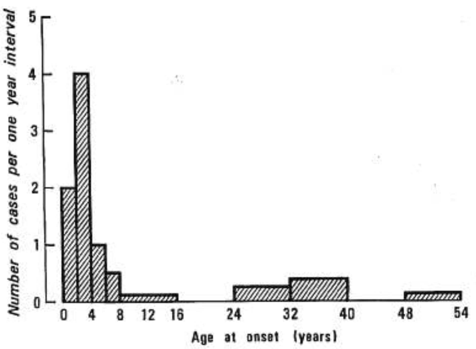
Figure : Histogram showing distribution of age of onset of hearing loss in patients with Langerhans cell histiocytosis, from McCaffrey and McDonald (McCaffrey and McDonald 1979). Note the bimodal distribution, with a large peak at 3 years, and a smaller peak in the 4th decade.
This bimodal distribution has also been shown in later studies, such as in the literature review by Saliba and Sidani which reported that the mean age at diagnosis of hearing loss is 3.5 years in children, and 35.5 years in adults (Saliba and Sidani 2009).
Genetics
Most cases of LCH do not have any clear genetic basis. A genome-wide association study suggested that a mutation in the SMAD6 gene conferred increased susceptibility to LCH (Peckham-Gregory et al. 2017).
Pathophysiological mechanism of disease
Langerhans cell histiocytosis is a “sporadic myeloid neoplasm characterized by clonal proliferation of CDIa+/CD207+ myeloid dendritic cells” (Galluzzi and Garavello 2025) that comprise a “subpopulation of dendritic histiocytes and are primarily located in the basal layer of the epidermis, where they act as tissue macrophages” (Saliba and Sidani 2009).
LCH can potentially cause audiologic and vestibular symptoms through both peripheral (end-organ) involvement and central nervous system involvement.
LCH involvement of the temporal bone can result in hearing loss. Most literature reports that hearing loss in LCH is more commonly conductive; Saliba and Sidani comment that, “LCH hearing loss is commonly conductive secondary to middle ear soft tissue infiltration, to ossicles erosion and to external auditory canal obstruction” (Saliba and Sidani 2009). Not all investigators reach the same conclusion; for example, Kaino and colleagues conducted a retrospective review of 317 cases of pediatric LCH with multisystem or multifocal bone involvement, and identified 9 patients with hearing loss, of whom none had purely conductive hearing loss (Kaino et al. 2026).
The literature review by Saliba and Sidani (Saliba and Sidani 2009) observes that when LCH affects the temporal bone, semicircular canal involvement (lateral > posterior > superior) overall is more common than cochlear involvement. This is shown in Figure 2 below.
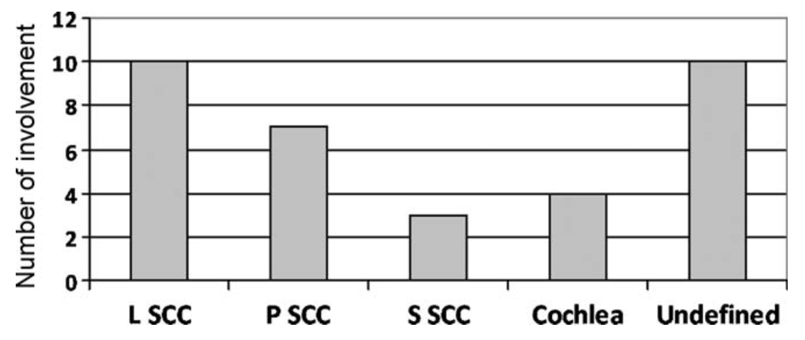
Figure : Otic capsule involvement in temporal bone Langerhans cell histiocytosis. Abbreviations: L=lateral; P=posterior; S=superior; SCC=semicircular canal. From Saliba and Sidani (Saliba and Sidani 2009).
LCH can also directly involve the central nervous system. Autier and colleagues (Autier et al. 2022) comment that:
“CNS impairment, termed neuro-LCH, is rare and occurs in 5 to 10% of cases. Two forms of neuro-LCH are described: (i) the tumor form and (ii) the degenerative form. The tumor form is characterized by proliferation of LCH cells within the CNS. In contrast, degenerative neuro-LCH (DN-LCH) is less well understood” (Autier et al. 2022).
Clinical presentation
The clinical presentation of LCH is quite variable. Galluzzi and Garavello (Galluzzi and Garavello 2025) comment:
“The clinical presentation is very heterogeneous and unspecific. Any organ or system of the body can be affected, but those more frequently involved are the skeleton (80% of cases), the skin (33%), and the pituitary (25%). Other organs involved are the liver, spleen, the hematopoietic system which includes bone marrow and lymph nodes (5–10%), the lungs (15% each), and the central nervous system (2–4%). The clinical course may vary from a self-limiting disease to a rapidly progressive that might lead to death” (Galluzzi and Garavello 2025).
As mentioned earlier, the mean age at diagnosis of hearing loss in LCH patients is 3.5 years in children, and 35.5 years in adults (Saliba and Sidani 2009).
The literature review by Saliba and Sidani (Saliba and Sidani 2009) found that hearing loss was reported in 7.4% – 28.5% of LCH patients; of all LCH patients with hearing loss, 10% were sensorineural; the remainder were conductive or mixed.
The presentation of the hearing loss is variable:
- Sudden sensorineural hearing loss, unilateral (Arsovic et al. 2013; Davoodi et al. 2026; Hore et al. 1999; Suzuki et al. 2010).
- Sensorineural hearing loss, bilateral and gradually progressive (Henderson et al. 2015).
- “1-year history of bilateral progressive hearing loss” (Gupta et al. 2018).
- “Progressive sensorineural or mixed hearing loss” (Mosnier et al. 2004).
Vestibular symptoms have also been reported in LCH (Achard et al. 2019; Achour et al. 2025; Amirdjazil et al. 1977; del Río et al. 2007; Losie et al. 2017; Nakamura et al. 2012; Nicholas and Garrahy 2019; Suzuki et al. 2010). The presentation of disequilibrium is variable:
- Some cases are abrupt in onset:
- “Gait instability over the past 5 days” (Nicholas and Garrahy 2019).
- “Sudden onset of… rotatory vertigo” (Suzuki et al. 2010).
- Other cases have a more gradual onset:
- “Progressive walking disturbances and recurrent falling” (Pähler Vor der Holte and Welkoborsky 2022).
- “Dizziness and unsteadiness that had been evolving for a year” (Achour et al. 2025).
- “Vertigo for 6 months” (Gupta et al. 2018).
- “Ataxia and a wide-based gait” (Nakamura et al. 2012).
Physical examination
Physical examination will depend on the organ(s) and site(s) of involvement. From an audiologic perspective, bedside physical examination (such as with a tuning fork) may show hearing loss that is unilateral, bilateral, sensorineural, conductive or mixed. From a vestibular standpoint, general neurological examination may show a wide-based gait, ataxia, or difficulty with unsighted tandem Romberg stance.
Ocular motor examination
Most reports do not document an ocular motor examination, or the examination is modest (e.g., “saccadic pursuit, a left hypermetria, a spontaneous up beating nystagmus, and a bilateral gaze nystagmus” (Achour et al. 2025)) or very limited (e.g., “There were 3 beats of end-point horizontal nystagmus noted with left gaze,” (Nicholas and Garrahy 2019)).
Testing: auditory
As described earlier, the hearing loss in LCH can be in any pattern: unilateral, bilateral, purely sensorineural, purely conductive, or mixed.
McCaffrey and McDonald (McCaffrey and McDonald 1979) provide an example of an LCH patient with left-sided temporal bone involvement who exhibited left-sided purely sensorineural hearing loss, more pronounced in the high frequencies. The corresponding audiogram is shown in Figure 3 below.
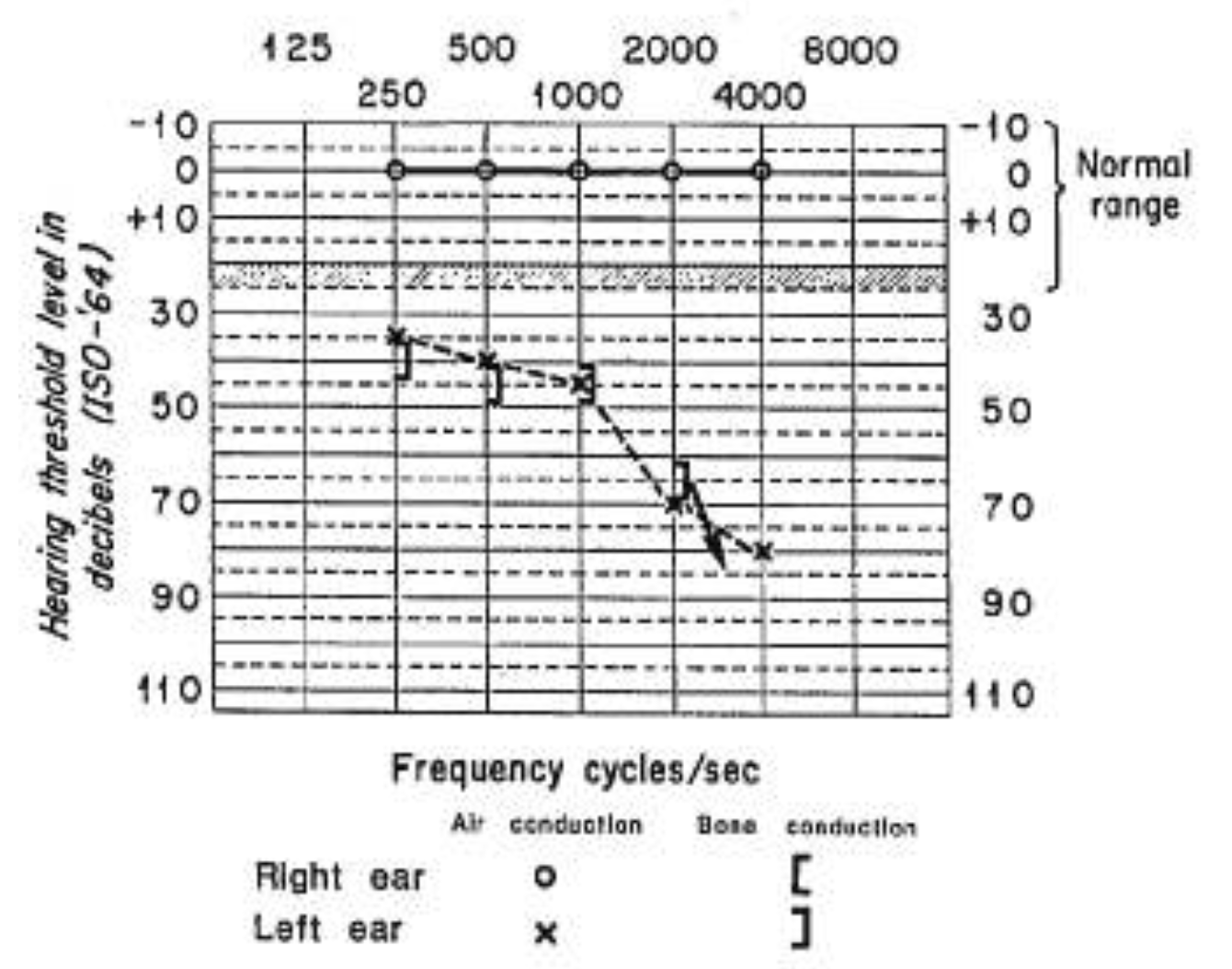
Figure : Audiometry from a patient with Langerhans cell histiocytosis involving the left temporal bone showing left-sided high greater than low frequency purely sensorineural hearing loss. From McCaffrey and McDonald (McCaffrey and McDonald 1979).
Pähler vor der Holte and Welkoborsky (Pähler Vor der Holte and Welkoborsky 2022) reported the case of a 4-year-old girl with LCH who presented with disequilibrium. Temporal bone CT showed asymmetrical temporal bone involvement, more pronounced on the right. Auditory brainstem evoked responses (ABR) were normal on the left, and abnormal on the right, as shown in Figure 4 below.
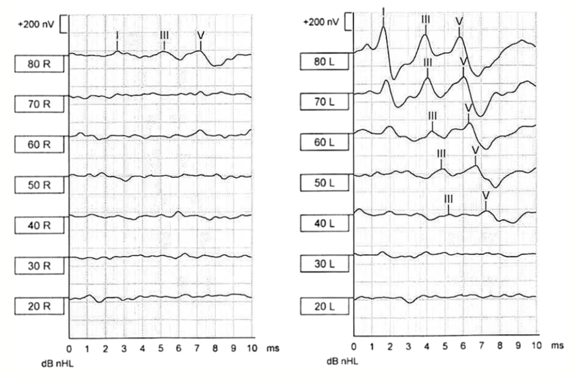
Figure : Pähler vor der Holte and Welkoborsky (Pähler Vor der Holte and Welkoborsky 2022) reported the case of a 4-year-old girl with right greater than left-sided temporal bone involvement by Langerhans cell histiocytosis. Brainstem auditory evoked responses (BAER) on the left ear (right side of figure) showed normal waveforms. BAER on the right side (left side of figure) showed a hearing threshold of 80 dB; the latency for wave I is markedly prolonged, with almost normal interpeak intervals, indicating a conductive or sensorineural hearing loss with intact vestibulocochlear nerve.
Testing: vestibular
Autier and colleagues (Autier et al. 2022) studied videonystagmography (VNG) in 20 LCH patients, and reported the prevalence of ocular motor abnormalities shown in Figure 5 below.
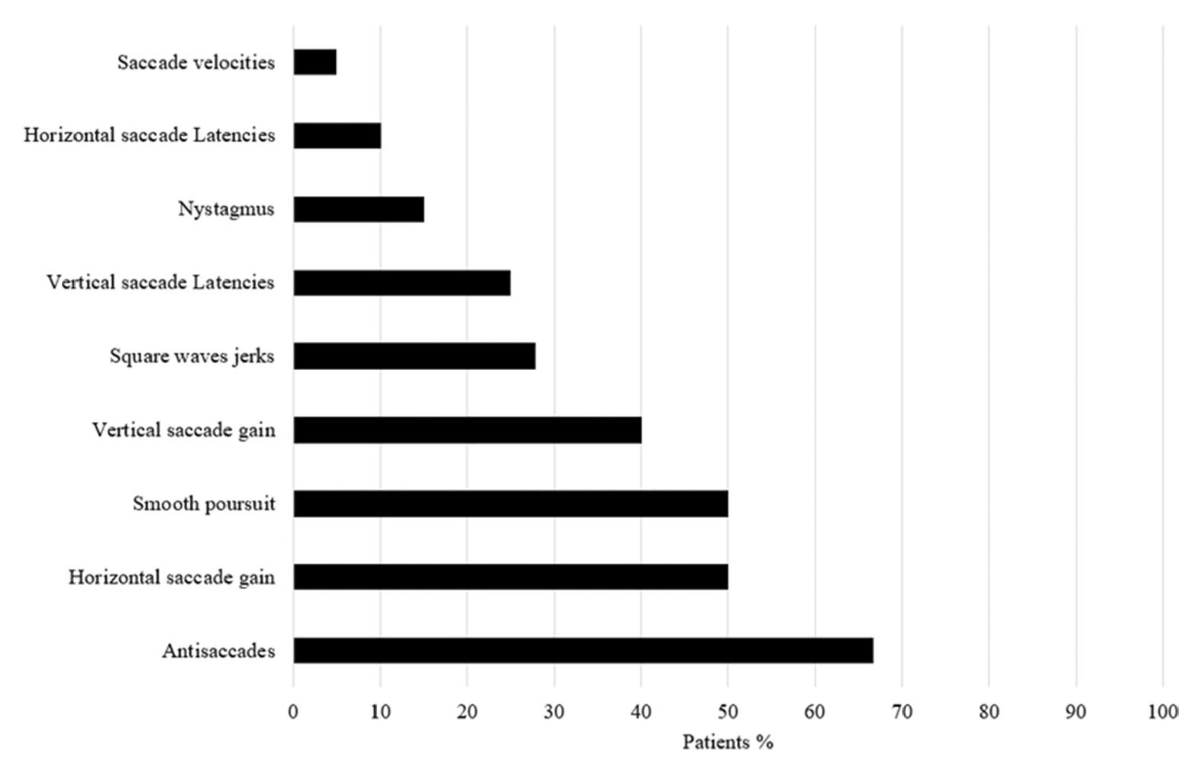
Figure : Prevalence of ocular motor abnormalities on videonystagmography (VNG) in 20 patients with Langerhans cell histiocytosis, as reported by Autier and colleagues (Autier et al. 2022).
Achour and colleagues (Achour et al. 2025) recorded the ocular motor findings of a 39-year-old lady with LCH and identified spontaneous up beat nystagmus, as well as post-headshake left beat nystagmus, as shown in Figure 6 below.
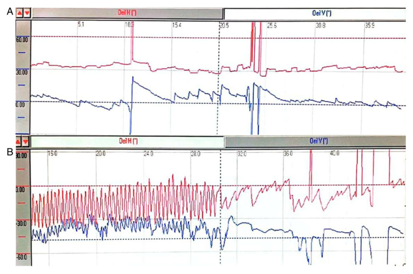
Figure : Ocular motor tracings from a 39-year-old lady with Langerhans cell histiocytosis. Panel A shows spontaneous up beat nystagmus. Panel B shows post-headshake left beat nystagmus. From Achour et al (Achour et al. 2025).
As of this writing there were no publications regarding LCH and other vestibular tests, such as cervical vestibular evoked myogenic potentials (cVEMP), ocular vestibular evoked myogenic potentials (oVEMP), video head impulse testing (vHIT), rotatory chair testing (RCT) or computerized dynamic posturography (CDP).
Imaging
In LCH patients with auditory and/or vestibular symptoms it is sensible to check both a temporal bone CT and an MRI of the brain and internal auditory canals without and with contrast, as each study provides different information. Examples are shown in Figure 7 and Figure 8.
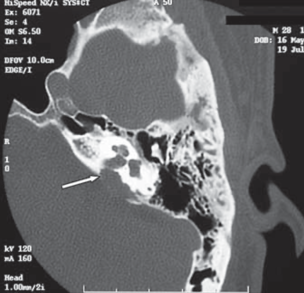
Figure : Axial CT of the temporal bone showing a lytic lesion of the posterior aspect of the left petrous bone, indicated by the white arrow. From del Rio et al (del Río et al. 2007).
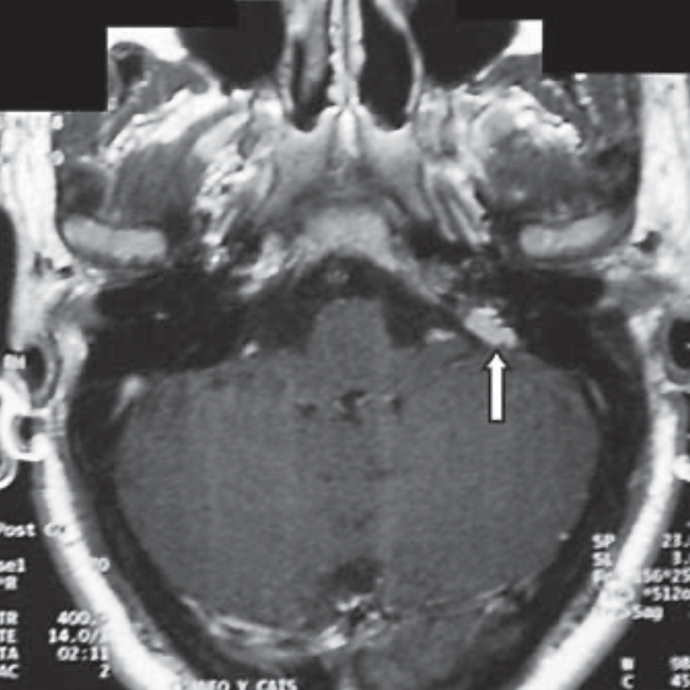
Figure : Axial T1 post-contrast MRI showing a gadolinium enhancing lesion in the left petrous bone surrounding the internal auditory canal, indicated by the white arrow. From del Rio et al (del Río et al. 2007).
Histopathology
Galluzzi and Garavello (Galluzzi and Garavello 2025) comment that:
“The definitive diagnosis of LCH is based on histological and immunophenotypic examination of lesional tissue. The main feature is the morphological identification of the characteristic LCH cells. Additionally, positive staining of the lesional cells with CD1a and/or Langerin (CD207) is required for definitive diagnosis” (Galluzzi and Garavello 2025).
Differential diagnosis
Given the variable presentation of audiologic and vestibular manifestations of LCH, the differential diagnosis is broad when considering only the clinical history, as it would include diseases that can cause any form of hearing loss, any type of disequilibrium, and their combination.
For example, the clinical history of an “isolated balance disorder” (Achard et al. 2019) is compatible with vestibular neuritis (VN) or an ataxia. The clinical history of “A 56-year-old woman complained of sudden onset of hearing loss on the left with rotatory vertigo” (Suzuki et al. 2010) is compatible with labyrinthitis, or a first attack of Ménière’s disease. The description of “a 30-year-old man presenting with vertigo and fluctuating hearing loss” (del Río et al. 2007) is compatible with Ménière’s disease.
Management
Management of LCH, particularly when there is temporal bone involvement, is typically a multidisciplinary undertaking, and may include otolaryngology, audiology, hematology-oncology, radiation oncology, physical therapy and others. Approaches include intralesional steroids, surgical excision, chemotherapy or radiation therapy.
Nanduri and colleagues (Nanduri et al. 1998) comment that, “The main role of surgery in [LCH] patients with ear involvement is (a) for performing diagnostic biopsy, and (b) to gain access for intralesional steroid injection.”
LCH involvement of the temporal bone is sometimes treated with radiation therapy (del Río et al. 2007; Zlodre et al. 2009).
For management of the hearing loss, in some cases cochlear implantation is considered, either unilateral (Gupta et al. 2018) or bilateral (Boyle et al. 2026; Segel and McKinnon 2013).
As of this writing there are no published studies regarding treatment of vestibular symptoms in LCH patients. If an LCH patient complains of disequilibrium, and especially if vestibular workup identifies a vestibular weakness, then a trial of vestibular rehabilitation therapy (VRT) is medical reasonable and low-risk.
Prognosis
Galluzzi and Garavello (Galluzzi and Garavello 2025) comment that:
“The prognosis depends on the number and extent of organ involvement. While survival for patients without organ dysfunction is excellent, mortality rates for patients with organ dysfunction may reach 20 %” (Galluzzi and Garavello 2025).
Prognosis of hearing loss is generally unfavorable, though partial recovery of hearing loss has been reported with chemotherapy (Losie et al. 2017) or radiation therapy (Hore et al. 1999).
Additional notes
Cases of LCH that initially manifest with purely aural symptoms (e.g., discharge) or purely auditory symptoms (e.g., hearing loss, otalgia) may first present to an otolaryngologist. Cases whose initial manifestation is isolated disequilibrium may first present to a physical therapist or neurologist.
References
Achard S, Parodi M, Denoyelle F (2019) Rare cause of vertigo in a child. Eur Ann Otorhinolaryngol Head Neck Dis 136: 227-228. doi: 10.1016/j.anorl.2019.04.009
Achour I, Kharrat I, Hbaieb Y, Ben Ayed M, Mnejja M, Hammami B, Souissi B, Charfeddine I (2025) Unusual Neurological Manifestation of Langerhans Cell Histiocytosis in an Adult. Ear Nose Throat J 104: 213-217. doi: 10.1177/01455613221106220
Amirdjazil Z, Esca SA, Konrad K (1977) Histiocytosis X in adult with skin and uncommon central nervous system involvement. Dermatologica 155: 283-91. doi: 10.1159/000250979
Arsovic N, Tomanovic N, Bukurov B (2013) Sudden sensorineural hearing loss as first presenting symptom of unifocal Langerhans cell histiocytosis in the temporal bone. Otol Neurotol 34: e24-5. doi: 10.1097/MAO.0b013e3182877b16
Autier L, Gaymard B, Bayen E, Del Cul A, Cohen-Aubart F, Martin-Duverneuil N, Haroche J, Mokhtari K, Héritier S, Donadieu J, Hoang-Xuan K, Idbaih A (2022) Eye movement abnormalities in neurodegenerative langerhans cell histiocytosis. Neurol Sci 43: 6539-6546. doi: 10.1007/s10072-022-06180-y
Baumann C, Reschke K, Jungehülsing M, Hierholzer J (2006) Destruction of the vestibular organ by Langerhans’ cell histiocytosis. Eur Radiol 16: 1177-8. doi: 10.1007/s00330-005-0024-1
Boyle S, Brennan M, Alshaikh K, Franklin CS, Bacon L, Flavin R, Glynn F, Walshe P, Gendre A (2026) Progressive cochlear ossification secondary to Langerhans cell histiocytosis requiring sequential bilateral cochlear implantation: a case report. Cochlear Implants Int 27: 247-252. doi: 10.1080/14670100.2026.2670091
Chu T, D’Angio GJ, Favara B, Ladisch S, Nesbit M, Pritchard J (1987) Histiocytosis syndromes in children. Writing Group of the Histiocyte Society. Lancet 1 8526: 208-9.
Davoodi F, Babamohamadi Z, Mokhtarinejad F, Khajavi M, Fallahi M, Abdoli Y (2026) A case of Langerhans cell histiocytosis localized in the temporal bone, which presented as sudden sensorineural hearing loss: A case report and review of the literature. SAGE Open Med Case Rep 14: 2050313×251411135. doi: 10.1177/2050313×251411135
del Río L, Lassaletta L, Martínez R, Sarriá MJ, Gavilán J (2007) Petrous bone Langerhans cell histiocytosis treated with radiosurgery. Stereotact Funct Neurosurg 85: 129-31. doi: 10.1159/000098528
Galluzzi F, Garavello W (2025) Temporal bone histiocytosis in children: a systematic review. Int J Pediatr Otorhinolaryngol 195: 112436. doi: 10.1016/j.ijporl.2025.112436
Goyal G, Shah MV, Hook CC, Wolanskyj AP, Call TG, Rech KL, Go RS (2018) Adult disseminated Langerhans cell histiocytosis: incidence, racial disparities and long-term outcomes. Br J Haematol 182: 579-581. doi: 10.1111/bjh.14818
Gupta G, Jain A, Grover M (2018) Successful cochlear implantation in Langerhans cell histiocytosis: A rare case. Cochlear Implants Int 19: 115-118. doi: 10.1080/14670100.2017.1402473
Henderson AH, Khan AA, Good C, Baer ST (2015) A case of bilateral sensorineural hearing loss from Langerhans cell histiocytosis. BMJ Case Rep 2015. doi: 10.1136/bcr-2015-210996
Hore I, Mitchell RB, Radcliffe G, Quiney R, Walker T (1999) Langerhans’ cell histiocytosis–a rare cause of sudden onset unilateral sensorineural hearing loss. J Laryngol Otol 113: 1098-100. doi: 10.1017/s0022215100157998
Kaino A, Sakamoto K, Moriya K, Osone S, Imamura T, Kudo K, Shioda Y, Imashuku S, Morimoto A (2026) Hearing loss in Langerhans cell histiocytosis: close association with central nervous system consequences. Int J Hematol 123: 916-923. doi: 10.1007/s12185-026-04160-y
Lichtenstein L (1953) Histiocytosis X; integration of eosinophilic granuloma of bone, Letterer-Siwe disease, and Schuller-Christian disease as related manifestations of a single nosologic entity. AMA Arch Pathol 56: 84-102.
Liu H, Stiller CA, Crooks CJ, Rous B, Bythell M, Broggio J, Rankin J, Nanduri V, Lanyon P, Card TR, Ban L, Elliss-Brookes L, Broughan JM, Paley L, Wong K, Bacon A, Bishton M, West J (2022) Incidence, prevalence and survival in patients with Langerhans cell histiocytosis: A national registry study from England, 2013-2019. Br J Haematol 199: 728-738. doi: 10.1111/bjh.18459
Losie JA, Yong M, Kozak FK, Chadha NK (2017) Unique Case of Hearing Recovery After Otic Capsule Destruction and Complete Sensorineural Hearing Loss Caused by Langerhans Cell Histiocytosis. Otol Neurotol 38: 1129-1132. doi: 10.1097/mao.0000000000001511
McCaffrey TV, McDonald TJ (1979) Histiocytosis X of the ear and temporal bone: review of 22 cases. Laryngoscope 89: 1735-42. doi: 10.1288/00005537-197911000-00004
Mosnier I, Rondini-Gilli E, Crosara PT, Belmatoug N, Cyna-Gorse F, Cazals-Hatem D, Abbey-Toby A, Bozorg-Grayeli A, Sterkers O (2004) Langerhans’ cell histiocytosis of the labyrinth in adults. Otol Neurotol 25: 27-32. doi: 10.1097/00129492-200401000-00006
Nakamura T, Morimoto N, Goto F, Shioda Y, Hoshino H, Kubota M, Taiji H (2012) Langerhans cell histiocytosis with disequilibrium. Auris Nasus Larynx 39: 627-30. doi: 10.1016/j.anl.2012.01.003
Nanduri VR, Pritchard J, Chong WK, Phelps PD, Sirimanna K, Bailey CM (1998) Labyrinthine involvement in Langerhans’ cell histiocytosis. Int J Pediatr Otorhinolaryngol 46: 109-15. doi: 10.1016/s0165-5876(98)00116-5
Nicholas PD, 3rd, Garrahy I (2019) A case of multisystem Langerhans cell histiocytosis presenting as central diabetes insipidus. J Community Hosp Intern Med Perspect 9: 515-517. doi: 10.1080/20009666.2019.1698231
Pähler Vor der Holte A, Welkoborsky HJ (2022) Case report: Langerhans cell histiocytosis of the temporal bone in children: Challenging diagnosis of a rare disease with some pitfalls. Clin Case Rep 10: e6057. doi: 10.1002/ccr3.6057
Peckham-Gregory EC, Chakraborty R, Scheurer ME, Belmont JW, Abhyankar H, Sengal AG, Scull BP, Eckstein O, Zinn DJ, Mayer L, Shih A, Merad M, Parsons DW, McClain KL, Lupo PJ, Allen CE (2017) A genome-wide association study of LCH identifies a variant in SMAD6 associated with susceptibility. Blood 130: 2229-2232. doi: 10.1182/blood-2017-08-800565
Saliba I, Sidani K (2009) Prognostic indicators for sensorineural hearing loss in temporal bone histiocytosis. Int J Pediatr Otorhinolaryngol 73: 1616-20. doi: 10.1016/j.ijporl.2009.07.010
Segel JM, McKinnon BJ (2013) Bilateral cochlear implantation in bilateral Langerhans cell histiocytosis. Cochlear Implants Int 14: 178-80. doi: 10.1179/1754762812y.0000000023
Suzuki H, Nishizawa S, Hohchi N, Wakasugi T, Shibata M, Ohkubo J, Tsukada J (2010) Langerhans cell histiocytosis of the petrous bone with sudden sensorineural hearing loss. Case report. Neurol Med Chir (Tokyo) 50: 693-7. doi: 10.2176/nmc.50.693
Zlodre JK, Rennie AT, Ramsden JD (2009) Langerhans’ cell histiocytosis of the temporal bone: successful treatment of sensorineural hearing loss with low-dose radiotherapy. J Laryngol Otol 123: 676-9. doi: 10.1017/s0022215108002995
