By Marcello Cherchi, MD PhD
For patients
In some cases when a doctor wants to look in your ear, they will want to use a special microscope with good illumination and magnification, called “binocular otomicroscopy.” During this procedure you are seated in a chair with a headrest. The doctor may place a small plastic or metal funnel into your ear canal, and look through it, to see your eardrum. If there is wax or a foreign object in your ear, this is a good technique for seeing it and (if possible) removing it. Some patients find it uncomfortable to have the small funnel in their ear. If the doctor is only looking in your ear, then this test may take only 10 – 20 seconds per ear. If the doctor is removing was from you ear, this may take several minutes. You do not need any special preparation for this test. There are no specific instructions for you to do after the test.
For clinicians
Overview
Binocular otomicroscopy involves using an operating microscope to examine the external auditory canal and tympanic membrane. Compared to handheld monocular otoscopy, binocular otomicroscopy provides several advantages, including better illumination, better magnification, adjustable focus, and stereopsis (and thus depth perception), and it frees the examiner’s hands to do other tasks (such as manipulate a speculum, disimpact ear wax, etc.).
Discussion
Binocular otomicroscopy refers to the use of a type of operating microscope to examine the ear. The otomicroscopes used today descend from the combined ingenuity of Carl Zeiss (1816 – 1888) and Carl Olof Siggesson Nylén (1892 – 1978) (Dohlman 1969).
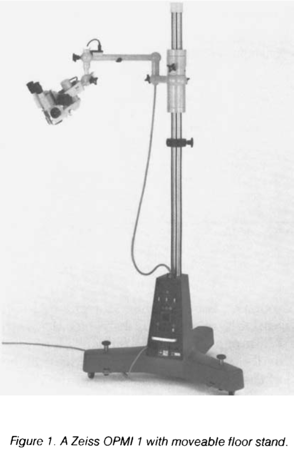
The Figure below, from Hoerenz (Hoerenz 1980), shows an old Carl Zeiss OPMI-1 operating microscope (no longer manufactured) that we used for many years, with a suspension system and floor stand.
The Figure below, from Hoerenz (Hoerenz 1980), depicts a schematic of the lens configuration for a binocular operating microscope.

For outpatient clinical purposes, a desirable range for the magnification changer is either 1 to 25x, or 1 to 40x.
The main objective lens of an operating microscope can be either fixed or variable. A fixed objective lens has an unvarying distance of best acuity between the lens and the subject; this configuration is adequate for outpatient clinical uses, and is less expensive; we prefer a fixed objective lens with a focal distance of 250 mm. A variable objective lens effectively provides “zoom” capabilities; this configuration is helpful when the microscope is deployed in an operating room; it is more expensive and generally unnecessary for outpatient clinical purposes.
Illumination for an operating microscope is provided by a coaxial system, as shown in the Figure below, from Hoerenz (Hoerenz 1980).

Most contemporary microscopes deliver illumination via a fiber optic cord. For many years the main type of illumination consisted of either incandescent or halogen bulbs; this is being gradually supplanted by LED illuminators, which are brighter and more expensive, but need replacement and maintenance less often.
In comparison to the handheld monocular otoscope, a binocular operating microscope offers several advantages:
- It frees the examiner’s hands, permitting instrumentation of the ear.
- The illumination source is strong.
- The magnification is good.
- The focus is adjustable.
- The binocularity permits stereopsis and thus depth perception, which is important when instrumenting the ear even for simple tasks (such as removing wax).
For these reasons, binocular otomicroscopy is superior to handheld monocular otoscopy, though the equipment is much more expensive. Our practice is to use binocular otomicroscopy whenever available; if that is unavailable, then we will use handheld monocular otoscopy.
Binocular otomicroscopy is helpful for identifying basic pathologies such as cerumen, middle ear effusions and tympanic membrane perforations. The magnification is sufficient that it can identify subtler findings, such as dimpling of the tympanic membrane in patients with tensor tympani myoclonus. The fact that it frees the examiner’s hands permits instrumentation for cerumen disimpaction or removal of foreign objects. The fact that it affords stereopsis and depth perception makes it safer to perform instrumented procedures.
During binocular otomicroscopy the patient is usually seated in an examination char with the head supported by a headrest so as to minimize movement. The examiner often uses an otologic speculum which facilitates visualizing the tympanic membrane.
References
Dohlman GF (1969) Carl Olof Nylen and the birth of the otomicroscope and microsurgery. Arch Otolaryngol 90: 813-7. doi: 10.1001/archotol.1969.00770030815025
Hoerenz P (1980) The operating microscope. I. Optical principles, illumination systems, and support systems. J Microsurg 1: 364-9. doi: 10.1002/micr.1920010506
![]()