By Marcello Cherchi, MD PhD
For patients
The phrase “Ehlers-Danlos syndrome” (EDS) refers to a group of diseases that can cause problems with joints, skin, blood vessels, the heart, the eyes and other body organs. It may be helpful to talk with a medical genetic counselor if you or your doctor think you might have EDS. Some EDS patients have hearing loss. Some EDS patients may feel disequilibrium because of changes in blood pressure, an inner ear problem, or nerve problems.
For clinicians
Overview
The term “Ehlers-Danlos syndrome (EDS)” refers to a collection of diseases resulting from mutations in specific genes that encode connective tissues or specific enzymes. These can manifest with a rather broad array of phenotypes. Genetic testing can confirm the diagnosis. Some patients with EDS have hearing loss of various types. Some EDS patients may experience disequilibrium, for which plausible proximal mechanisms include orthostatic intolerance, superior semicircular canal dehiscence and peripheral neuropathy.
Introduction
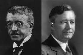
The first cases of what came to be known as Ehlers-Danlos syndrome (EDS) were originally described by Danish dermatologist, Dr. Edvard Lauritz Ehlers (1863 – 1937) (Ehlers 1901) and French dermatologist, Henri-Alexandre Danlos (1844 – 1912) (Danlos 1908). The eponymous designation of “Ehlers-Danlos syndrome” appears to have been originally proposed in 1936 by the English physician, Dr. Frederick Parkes Weber (Weber 1936).
As additional cases emerged, it became apparent that there was significant phenotypic variability. With the advent of genetics, it is now understood that EDS comprises a range of distinct pathologies with different, though overlapping, clinical manifestations. The Table below summarizes the types of Ehlers-Danlos syndrome in Online Mendelian Inheritance in Man as of this writing.
|
Type |
Other nomenclature |
Inheritance pattern |
OMIM disease |
OMIM gene |
Cytogenetic location |
Gene(s) |
Protein |
|---|---|---|---|---|---|---|---|
|
Classical EDS, type 1 |
cEDS, EDS type I |
AD |
9q34.3 |
COL5A1 |
Type V collagen, alpha 1 |
||
|
Classical EDS, type 1 |
cEDS, EDS type I |
AD |
17q21.33 |
COL1A1 c.934C>T, p.(Arg312Cus) |
Type I collagen, alpha 1 |
||
|
Classical EDS, type 2 |
cEDS, type II |
2q32.2 |
COL5A2 |
Type 5 collagen, alpha 2 |
|||
|
Hypermobile type EDS |
clEDS, EDS type III |
AD |
6p21.3 |
TNXB |
Tenascin XB, isoform 1 |
||
|
Classic-like EDS |
EDS with TNXB deficiency |
AR |
6p21.3 |
TNXB |
Tenascin XB, isoform 1 |
||
|
Vascular EDS |
EDS type IV |
AD |
2q32.2 |
COL3A1 |
Type III collagen, alpha 1 |
||
|
Kyphoscoliotic EDS, type 1 |
EDS KSCL1, EDS type VIA |
AR |
1p36.22 |
• PLOD1 (procollagen-lysine, 2-oxoglutarate 5-dioxygenase) |
PLOD1 (procollagen-lysine, 2-oxoglutarate 5-dioxygenase) |
||
|
Kyphoscoliotic EDS, type 2 |
EDS type VIB |
AR |
7p14.3 |
FKBP14 |
FK506 binding protein 14 |
||
|
Brittle cornea syndrome, type 1 |
BCS1 |
AR |
16q24.2 |
ZNF469 (zinc finger protein 469) |
ZNF469 (zinc finger protein 469) |
||
|
Brittle cornea syndrome, type 2 |
BCS2 |
AR |
4q27 |
PRDM5 |
PRDM5 (PR domain-containing protein 5) |
||
|
Musculocontractural EDS type 1 |
mcEDS, D4ST1-deficient type EDS, EDS type VIB |
AR |
15q15.1 |
CHST14 |
CHST14 (carbohydrate sulfotransferase 14) |
||
|
Arthrochalasic EDS type 1 |
aEDS, EDS type VIIA |
AD |
17q21.33 |
COL1A1 |
Type I collagen, alpha 1 |
||
|
Arthrochalasic EDS type 2 |
aEDS, EDS type VIIB |
AD |
7q21.3 |
COL1A2 |
Type I collagen, alpha 2 |
||
|
Cardiac-valvular EDS |
cvEDS, EDS with COL1A2 deficiency |
AR |
7q21.3 |
COL1A2 (biallelic mutations that lead to COL1A2 NMD and absence of pro a2(I) collagen chains) |
Type I collagen, alpha 2 |
||
|
Dermatosparexis EDS |
EDS type VIIC |
AR |
5q35.3 |
ADAMTS2 |
A disintegrin-like and metalloproteinase with thrombospondin type 1 motif 2 |
||
|
Spondylodysplastic EDS, type 1 |
EDS SPD1 |
AR |
5q35.3 |
B4GALT7 |
Beta-1,4-galactosyltransferase 7 |
||
|
Spondylodysplastic EDS, type 2 |
EDS SPD2 |
AR |
1p36.33 |
B3GALT6 |
Beta-1,3-galactosyltransferase 6 |
||
|
EDS, spondylocheirodysplastic form, type 3 |
SCD-EDS |
AR |
11p11.2 |
SLC39A13 |
Solute carrier family 39 (zinc transporter), member 13 |
||
|
Periodontal EDS |
EDS PD1 |
AD |
12p13.31 |
C1R |
Complement component 1, r subcomponent |
||
|
Vascular EDS |
EDS VASC |
AD |
2q32.2 |
COL3A1 |
Collagen type III, alpha 1 |
||
|
EDS with platelet dysfunction from fibronectin abnormality |
EDS type X |
AD |
2q35 |
FN1 |
Fibronectin |
Table : Types of Ehlers-Danlos syndrome. AD = autosomal dominant, AR = autosomal recessive, OMIM = Online Mendelian Inheritance in Man
Genetics
Most subtypes of Ehlers-Danlos syndrome result from mutations in genes encoding proteins for connective tissue structures or for enzymes.
Pathophysiological mechanism of disease: hearing loss
Weir and colleagues studied audiometry in 141 pediatric patients diagnosed with EDS, and found some form of hearing loss in 32 (22.7%). Of those 32 patients, the hearing loss was bilateral in 19 (59%) and unilateral in 13 (41%). Out of 282 ears (in the 141 EDS patients), pure conductive hearing loss was found in 25 (8.5% of ears), pure sensorineural hearing loss was found in 23 (8.2% of ears), and mixed hearing loss was found in only 3 (1.4% of ears). The authors reported that of all the EDS patients with hearing loss, 50% “received more than two diagnoses of otitis media throughout the course of their care,” and “31.4% of the cohort with hearing loss had a diagnosis of Eustachian tube dysfunction.” There are additionally several case reports of otosclerosis in EDS patients (Miyajima, Ishimoto, Yamasoba 2007; Thomas et al. 1996).
Pathophysiological mechanism of disease: disequilibrium
As far as symptoms of disequilibrium are concerned, there are several potential mechanisms by which EDS can cause disorders of equilibrium.
The most common mechanism appears to be autonomic dysfunction, manifesting primarily as orthostatic intolerance, particularly postural orthostatic tachycardia syndrome (POTS) (Celletti et al. 2020; Celletti et al. 2017; De Wandele et al. 2014; Hakim et al. 2017; Mathias et al. 2021; Miller et al. 2020; Qarajeh et al. 2021; Roma et al. 2018; Rowe et al. 1999; Wallman, Weinberg, Hohler 2014).
Superior semicircular canal dehiscence (SSCD) has been reported in several cases of EDS, including unilateral (Chung et al. 2017) and bilateral (Preet et al. 2019; Unterberger et al. 2021).
Peripheral neuropathy of various types may also be contributory (Cazzato et al. 2016; Cook and Jordan 2021; Fernandez et al. 2022; Galan and Kousseff 1995; Igharo et al. 2023; Pascarella et al. 2016; Schady and Ochoa 1984; Voermans et al. 2006; Voermans, Knoop, van Engelen 2011).
In EDS patients with altered cervical mechanics, cervicogenic vertigo (CV) is also a reasonable consideration, though this has not been formally studied.
Clinical presentation
The clinical presentation depends on the proximal mechanism of the symptom of disequilibrium.
Physical examination
Physical examination findings will depend on the proximal mechanism of the symptom of disequilibrium.
It is reasonable for physical examination to include comparison of supine and standing pulse/pressure to seek evidence of postural orthostatic tachycardia.
Ocular motor examination
It is reasonable on infrared video oculography to assess for Valsalva-induced nystagmus which may be elicit nystagmus if superior semicircular canal dehiscence is present.
Testing: audiologic
If an EDS patient has hearing complaints it is reasonable to check audiometry.
Testing: vestibular
Consider checking audiometry, cervical vestibular evoked myogenic potentials and ocular vestibular evoked myogenic potentials to evaluate for superior semicircular canal dehiscence.
Consider checking computerized dynamic posturography for an overall quantification of balance.
Testing: other
Consider checking a tilt table test to evaluate for evidence of autonomic dysfunction.
Consider consultation with a medical geneticist regarding whether genetic testing is appropriate and, if so, which variants should be checked.
Imaging
From the otoneurological perspective, if screening tests (audiometry, cervical vestibular evoked myogenic potentials, ocular vestibular evoked myogenic potentials) are compatible with the presence superior semicircular canal dehiscence, then consider a temporal bone CT without contrast.
Treatment
Care of an EDS patient tends to be multi-disciplinary (Cherchi 2026).
If a patient or their doctor suspects EDS, then consultation with a medical geneticist is sensible to determine whether genetic testing for diagnostic confirmation is appropriate.
Depending on the genetic subtype and clinical manifestations, the overall care of an EDS patient is often organized by a rheumatologist, often in consultation with other specialists, such as a dermatologist.
In EDS patients with hearing loss, referral to audiology is reasonable.
In EDS patients with superior semicircular canal dehiscence, referral to otolaryngology is reasonable. Unfortunately, EDS patients are often poor surgical candidates due to poor tissue healing and in some cases vascular complications.
In EDS patients with postural orthostatic tachycardia syndrome, referral to cardiology is reasonable.
References
Cazzato D, Castori M, Lombardi R, Caravello F, Bella ED, Petrucci A, Grammatico P, Dordoni C, Colombi M, Lauria G (2016) Small fiber neuropathy is a common feature of Ehlers-Danlos syndromes. Neurology 87: 155-9. doi: 10.1212/WNL.0000000000002847
Celletti C, Borsellino B, Castori M, Censi F, Calcagnini G, Camerota F, Strano S (2020) A new insight on postural tachycardia syndrome in 102 adults with hypermobile Ehlers-Danlos Syndrome/hypermobility spectrum disorder. Monaldi Arch Chest Dis 90. doi: 10.4081/monaldi.2020.1286
Celletti C, Camerota F, Castori M, Censi F, Gioffre L, Calcagnini G, Strano S (2017) Orthostatic Intolerance and Postural Orthostatic Tachycardia Syndrome in Joint Hypermobility Syndrome/Ehlers-Danlos Syndrome, Hypermobility Type: Neurovegetative Dysregulation or Autonomic Failure? Biomed Res Int 2017: 9161865. doi: 10.1155/2017/9161865
Cherchi M (2026) Audio-vestibular manifestations of Ehlers-Danlos syndrome. Future Rare Diseases 6: 2701706. doi: 10.1080/23995270.2026.2701706
Chung LK, Lagman C, Nagasawa DT, Gopen Q, Yang I (2017) Superior Semicircular Canal Dehiscence in a Patient with Ehlers-Danlos Syndrome: A Case Report. Cureus 9: e1141. doi: 10.7759/cureus.1141
Cook MK, Jordan M (2021) Autoimmune Small Fiber Neuropathy Associated With Ehlers-Danlos Syndrome Treated With Intravenous Immunoglobulins. J Clin Neuromuscul Dis 22: 160-163. doi: 10.1097/CND.0000000000000341
Danlos H-A (1908) Un cas de cutis laxa avec tumeurs par comtusion chronique des coudes et des genoux (xanthome juvénile pseudo-diabetique de MM Hallopeau et Macé de Lépinay) [A case of cutis laxa with chronic comtusion tumors of the elbows and knees (pseudo-diabetic juvenile xanthoma of MM Hallopeau and Macé de Lépinay)]. Bulletin de la Societé francaise de dermatologie et de syphiligraphie 19: 70-72.
De Wandele I, Rombaut L, Leybaert L, Van de Borne P, De Backer T, Malfait F, De Paepe A, Calders P (2014) Dysautonomia and its underlying mechanisms in the hypermobility type of Ehlers-Danlos syndrome. Semin Arthritis Rheum 44: 93-100. doi: 10.1016/j.semarthrit.2013.12.006
Ehlers EL (1901) Cutis laxa. Neigung zu Haemorrhagien in der Haut, Lockering mehrerer Artikulationen [Lax skin: Tendency to hemorrhages in the skin, loosening of several articulations]. Dermatologische Zeitschrift 8: 173-174.
Fernandez A, Aubry-Rozier B, Vautey M, Berna C, Suter MR (2022) Small fiber neuropathy in hypermobile Ehlers Danlos syndrome/hypermobility spectrum disorder. J Intern Med 292: 957-960. doi: 10.1111/joim.13539
Galan E, Kousseff BG (1995) Peripheral neuropathy in Ehlers-Danlos syndrome. Pediatr Neurol 12: 242-5. doi: 10.1016/0887-8994(95)00003-x
Hakim A, O’Callaghan C, De Wandele I, Stiles L, Pocinki A, Rowe P (2017) Cardiovascular autonomic dysfunction in Ehlers-Danlos syndrome-Hypermobile type. Am J Med Genet C Semin Med Genet 175: 168-174. doi: 10.1002/ajmg.c.31543
Igharo D, Thiel JC, Rolke R, Akkaya M, Weis J, Katona I, Schulz JB, Maier A (2023) Skin biopsy reveals generalized small fibre neuropathy in hypermobile Ehlers-Danlos syndromes. Eur J Neurol 30: 719-728. doi: 10.1111/ene.15649
Mathias CJ, Owens A, Iodice V, Hakim A (2021) Dysautonomia in the Ehlers-Danlos syndromes and hypermobility spectrum disorders-With a focus on the postural tachycardia syndrome. Am J Med Genet C Semin Med Genet 187: 510-519. doi: 10.1002/ajmg.c.31951
Miller AJ, Stiles LE, Sheehan T, Bascom R, Levy HP, Francomano CA, Arnold AC (2020) Prevalence of hypermobile Ehlers-Danlos syndrome in postural orthostatic tachycardia syndrome. Auton Neurosci 224: 102637. doi: 10.1016/j.autneu.2020.102637
Miyajima C, Ishimoto S, Yamasoba T (2007) Otosclerosis associated with Ehlers-Danlos syndrome: report of a case. Acta Otolaryngol Suppl: 157-9. doi: 10.1080/03655230701600418
Pascarella A, Provitera V, Lullo F, Stancanelli A, Saltalamacchia AM, Caporaso G, Nolano M (2016) Evidence of small fiber neuropathy in a patient with Ehlers-Danlos syndrome, hypermobility-type. Clin Neurophysiol 127: 1914-6. doi: 10.1016/j.clinph.2015.12.004
Preet K, Udawatta M, Duong C, Gopen Q, Yang I (2019) Bilateral Superior Semicircular Canal Dehiscence Associated with Ehlers-Danlos Syndrome: A Report of 2 Cases. World Neurosurg 122: 161-164. doi: 10.1016/j.wneu.2018.10.126
Qarajeh R, Derbas LA, Abdallah M, Kureshi F (2021) A 21-year-old woman with Ehlers-Danlos syndrome and persistent lightheadedness. Cleve Clin J Med 88: 93-97. doi: 10.3949/ccjm.88a.19137
Roma M, Marden CL, De Wandele I, Francomano CA, Rowe PC (2018) Postural tachycardia syndrome and other forms of orthostatic intolerance in Ehlers-Danlos syndrome. Auton Neurosci 215: 89-96. doi: 10.1016/j.autneu.2018.02.006
Rowe PC, Barron DF, Calkins H, Maumenee IH, Tong PY, Geraghty MT (1999) Orthostatic intolerance and chronic fatigue syndrome associated with Ehlers-Danlos syndrome. J Pediatr 135: 494-9.
Schady W, Ochoa J (1984) Ehlers-Danlos in association with tomaculous neuropathy. Neurology 34: 1270-1. doi: 10.1212/wnl.34.9.1270-a
Thomas DM, Wright JL, Soucek S, Shalom AS (1996) Ehlers-Danlos syndrome: aural manifestations and treatment. Am J Otolaryngol 17: 432-3. doi: 10.1016/s0196-0709(96)90081-2
Unterberger A, Miller J, Gopen Q, Yang I (2021) Bilateral Superior Semicircular Canal Dehiscence Concurrent With Ehlers-Danlos Syndrome: A Case Report. Cureus 13: e19943. doi: 10.7759/cureus.19943
Voermans NC, Drost G, van Kampen A, Gabreels-Festen AA, Lammens M, Hamel BC, Schalkwijk J, van Engelen BG (2006) Recurrent neuropathy associated with Ehlers-Danlos syndrome. J Neurol 253: 670-1. doi: 10.1007/s00415-005-0056-0
Voermans NC, Knoop H, van Engelen BG (2011) High frequency of neuropathic pain in Ehlers-Danlos syndrome: an association with axonal polyneuropathy and compression neuropathy? J Pain Symptom Manage 41: e4-6; author reply e6-7. doi: 10.1016/j.jpainsymman.2011.02.006
Wallman D, Weinberg J, Hohler AD (2014) Ehlers-Danlos Syndrome and Postural Tachycardia Syndrome: a relationship study. J Neurol Sci 340: 99-102. doi: 10.1016/j.jns.2014.03.002
Weber FP (1936) Ehlers-Danlos Syndrome. Proceedings of the Royal Society of Medicine 30: 30-31. doi: 10.1177/003591573603000107
![]()